. 2015 Jun 17:14:120.
doi: 10.1186/s12943-015-0387-0.

A non-tight junction function of claudin-7-Interaction with integrin signaling in suppressing lung cancer cell proliferation and detachment

Affiliations

A non-tight junction function of claudin-7-Interaction with integrin signaling in suppressing lung cancer cell proliferation and detachment

Zhe Lu et al. Mol Cancer. .

Abstract

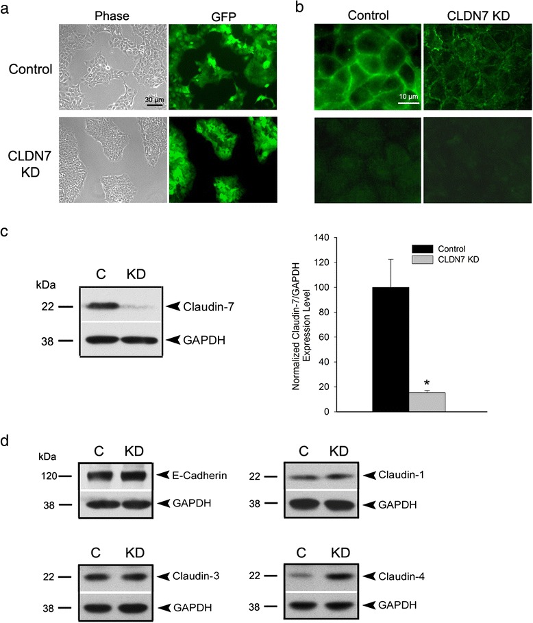
Background: Claudins are a family of tight junction (TJ) membrane proteins involved in a broad spectrum of human diseases including cancer. Claudin-7 is a unique TJ membrane protein in that it has a strong basolateral membrane distribution in epithelial cells and in tissues. Therefore, this study aims to investigate the functional significance of this non-TJ localization of claudin-7 in human lung cancer cells.

Methods: Claudin-7 expression was suppressed or deleted by lentivirus shRNA or by targeted-gene deletion. Cell cycle analysis and antibody blocking methods were employed to assay cell proliferation and cell attachment, respectively. Electron microscopy and transepthelial electrical resistance measurement were performed to examine the TJ ultrastructure and barrier function. Co-immunolocalization and co-immunoprecipitation was used to study claudin-7 interaction with integrin β1. Tumor growth in vivo were analyzed using athymic nude mice.

Results: Claudin-7 co-localizes and forms a stable complex with integrin β1. Both suppressing claudin-7 expression by lentivirus shRNA in human lung cancer cells (KD cells) and deletion of claudin-7 in mouse lungs lead to the reduction in integrin β1 and phospho-FAK levels. Suppressing claudin-7 expression increases cell growth and cell cycle progression. More significantly, claudin-7 KD cells have severe defects in cell-matrix interactions and adhere poorly to culture plates with a remarkably reduced integrin β1 expression. When cultured on uncoated glass coverslips, claudin-7 KD cells grow on top of each other and form spheroids while the control cells adhere well and grow as a monolayer. Reintroducing claudin-7 reduces cell proliferation, upregulates integrin β1 expression and increases cell-matrix adhesion. Integrin β1 transfection partially rescues the cell attachment defect. When inoculated into nude mice, claudin-7 KD cells produced significantly larger tumors than control cells.

Conclusion: In this study, we identified a previously unrecognized function of claudin-7 in regulating cell proliferation and maintaining epithelial cell attachment through engaging integrin β1.


Figures

Fig. 1
Fig. 1
Knockdown of claudin-7 in HCC827 lung cancer cells. a Representative phase and green fluorescence images of live control and claudin-7 KD cells. Both control and claudin-7 shRNA lentivirus constructs contain a GFP expression sequence. b Top panel is the anti-claudin-7 immunofluorescence staining of control and claudin-7 KD cells. Claudin-7 level was dramatically decreased in claudin-7 KD cells. The bottom panel is the anti-GFP immunofluorescence staining. The cells were fixed with 100 % methanol, which lead to GFP proteins leaking out of the cells so that the green fluorescence shown in the top panel was only the claudin-7 signal. c Western blot shows the diminished level of claudin-7 in the KD cells (left). The statistical analysis of claudin-7 expression in control and KD cells from five independent experiments is shown on right. *P < 0.05. d Protein expression levels of E-cadherin, claudin-1, −3, and −4 in control and KD cells. Only claudin-4 expression level was increased in KD cells. GAPDH served as a loading control
Fig. 2
Fig. 2
Increased cell proliferation in claudin-7 KD cells. a Five × 103 control and claudin-7 KD cells were seeded into 24-well plates and the cell number was counted for each sample on days 2, 4, and 6 after plating. Claudin-7 KD cells show significantly higher proliferation rate compared to the control cells on days 4 and 6. *P < 0.05. b Three days after plating, cells were trypsinized, centrifuged, and stained with propidium iodide and subjected to cell cycle analysis by flow cytometry. Claudin-7 KD cells show a higher percentage of G2/M and S phase cells and lower percentage of apoptotic cells. *P < 0.05. c Representative western blots show increased levels of phospho-ERK1/2, survivin, and phospho-Bcl-2 and decreased level of cleaved PARP in claudin-7 KD cells when compared to control cells. A total of 30 μg proteins were loaded for each sample. d The top panel is the representative cell cycle analysis of control and claudin-7 KD cells in regular culture medium 3 days after plating. The bottom panel was taken after 24 h of aphidicolin treatment, showing that both cell lines were inhibited at S phase. e Control and KD cells were treated without (left) and with 24 h of aphidicolin (right). Cell lysates were collected 2.5 and 18 h after releasing from aphidicolin treatment
Fig. 3
Fig. 3
Reduced cell-matrix adhesion in claudin-7 KD cells. a Left panel: Scratches were made on the confluent control and claudin-7 KD cell monolayer. Claudin-7 KD cells were easily peeled off along the scratch as shown in arrows, while the control cells were well attached to the plate. Right panel: When cultured on uncoated glass coverslips, claudin-7 KD formed spheroids while the control cells were able to spread out and formed a monolayer. b Monolayer cultures of control and claudin-7 KD cells were exposed to trypsinization and phase images were taken every minute. Claudin-7 KD cells took less time (4 min) to be fully lifted by trypsin than the control cells (7 min). c Cell attachment assay. Two × 105 control and KD cells were plated to each well of the collagen IV-coated 24-well plates. Four hours later, the unattached cells were washed off and the attached cells were trypsinized and counted. KD cells showed significantly reduced cell attachment compared to the control cells (left). The significantly reduced cell attachment was also observed in T-47D breast cancer cells with claudin-7 knockdown by the same shRNA approach (right). *P < 0.05. The insert shows the knockdown level of claudin-7 in T-47D cells. d Electron microscopy shows a larger gap between claudin-7 KD cells and the coverslip than that between control cells and the coverslip. TJs in both cell lines were largely intact (arrows). Magnifications: top, ×25,000; bottom, ×50,000. e TER measurement indicates that there was no significant change in the barrier function of TJs after claudin-7 was knocked down
Fig. 4
Fig. 4
Decreased mRNA and protein levels and disrupted localization of integrin β1 in claudin-7 KD cells. a Real-time RT-PCR was performed on integrins β1, β2, and β3. The mRNA level of integrin β1 was significantly reduced in claudin-7 KD cells. *P < 0.05. b Western blots show that integrin β1 and phospho-FAK levels were decreased in both claudin-7 KD cells (left) and claudin-7 knockout mouse lungs (right). At least six independent experiments were performed. c Immunofluorescence staining shows the localization of claudin-7 and integrin β1 in control and KD cells. Z-stack shows the co-localization of claudin-7 with integrin β1 at the basolateral surface in the merged image (top). In contrast, the localization of integrin β1 was greatly disrupted in the claudin-7 KD cells; it was no longer at the cell membrane (bottom). Arrows in control cells indicate the partial co-localization of claudin-7 with integrin β1. d Claudin-7 co-immunoprecipitated with integrin β1. Control cells were lysed in RIPA buffer without SDS and immunoprecipitated with either integrin β1 or claudin-7 antibody and then probed with either claudin-7 or integrin β1. e Control and KD cells were treated with anti-integrin β1 adhesion-blocking antibody or mouse IgG as a control. Cells were treated with the antibodies for two days, then cells were trypsinized and replated. The unattached cells were washed away two hours after replating, and the cells attached to the culture plate were trypsinized and counted. P values were shown above the bars on the right
Fig. 5
Fig. 5
Treatments of claudin-7 KD cells with collagens, genistein and aphidicolin. a Real-time RT-PCR revealed that several collagens were significantly decreased in claudin-7 KD cells when compared to the control cells. *P < 0.05. b By culturing the cells on type IV collagen-coated culture plates, claudin-7 KD cells displayed the increased cell size and were more spread out. c Five × 103 control or claudin-7 KD cells were plated on type IV collagen-coated culture plates. Cell number was counted on days 2, 4, and 6 after plating. Claudin-7 KD cells showed a higher proliferation rate than control cells. *P < 0.05. d Phase images of control and KD cells after 24 h of DMSO (control) or 200 μM genistein treatment. e Phase images of control and KD cells after 24 h of DMSO or 5 μg/ml aphidicolin treatment. f Control and KD cells cultured on the collagen IV coated plate, or treated with genistein or aphidicolin were lysed, loaded onto SDS-polyacrylamide gel, and probed with anti-integrin β1 antibody
Fig. 6
Fig. 6
Rescued claudin-7 KD cell defects by exogenous expression of claudin-7 or integrin β1 in KD cells. a The claudin-7 construct tagged with myc was transfected into the HCC827 claudin-7 KD cells. The transfected claudin-7 sequence has three nucleotide differences from lentivirus claudin-7 shRNA (due to mouse and human claudin-7 difference), so it is resistant to the lentivirus shRNA. The claudin-7 KD cells with claudin-7-myc transfection became larger in size and more spread out (phase images). Anti-myc immunofluorescence staining indicated by arrows shows the exogenous claudin-7 expression in claudin-7 KD cells. b Lysates from claudin-7 KD cells transfected with vector (KD + V) or claudin-7-myc (KD + 7) were collected and subjected to western blot analysis. The expression levels of integrin β1 and cleaved PARP were both increased in claudin-7 transfected KD cells. The membrane probed with anti-myc antibody shows the exogenous claudin-7 expression in KD cells. c Five × 103 claudin-7 KD cells transfected with vector or claudin-7-myc were plated into 24-well plates. Cell number was counted on days 1, 3, 5 after plating. *P < 0.05. d Claudin-7 KD cells transfected claudin-7 (upper panel) or integrin β1 (lower panel) construct were trypsinized and plated on uncoated glass coverslips. e Lysates from claudin-7 KD cells transfected with vector (KD + V) or integrin β1 (KD+ β1) were collected and subjected to western blot analysis. The membrane probed with anti-integrin β1 antibody shows the expression level of integrin β1 in KD cells without or with integrin β1 transfection
Fig. 7
Fig. 7
The inhibitory effect of claudin-7 on tumor growth in nude mice. a Two × 106 HCC827 control or claudin-7 KD cells were harvested and suspended in the culture medium, then subcutaneously injected into the left and right flanks of a total of 10 nude mice, respectively. Arrows indicate the tumors generated subcutaneously in the representative nude mouse which was sacrificed 30 days post tumor cell inoculation. b Western blot analysis shows the increased expression levels of phospho-ERK1/2, survivin, and cleaved PARP in claudin-7 KD cells-induced tumors when compared to those of control cells-induced tumors. c, d The nude mice were sacrificed and the tumors were removed and measured in both volume and weight. Statistical analysis was conducted on a total of 10 nude mice. Medians were shown as black solid lines in the figures. *P < 0.05. e, f Growth curves for ten control cells-and ten claudin-7 KD cells-inoculated tumors

References

    1. Stella GM, Luisetti M, Inghilleri S, Cemmi F, Scabini R, Zorzetto M, et al. Targeting EGFR in non-small-cell lung cancer: lessons, experiences, strategies. Respir Med. 2012;106:173–83. doi: 10.1016/j.rmed.2011.10.015. - DOI - PubMed
    1. Schneeberger EE, Lynch RD. The tight junction: a multifunctional complex. Am J Physiol Cell Physiol. 2004;286:C1213–28. doi: 10.1152/ajpcell.00558.2003. - DOI - PubMed
    1. Singh AB, Sharma A, Dhawan P. Claudin family of proteins and cancer: an overview. J Oncol. 2010;2010:541957. doi: 10.1155/2010/541957. - DOI - PMC - PubMed
    1. Benjamin JM, Nelson WJ. Bench to bedside and back again: molecular mechanisms of alpha-catenin function and roles in tumorigenesis. Semin Cancer Biol. 2008;18:53–64. doi: 10.1016/j.semcancer.2007.08.003. - DOI - PMC - PubMed
    1. Tsukita S, Furuse M, Itoh M. Multifunctional strands in tight junctions. Nat Rev Mol Cell Biol. 2001;2:285–93. doi: 10.1038/35067088. - DOI - PubMed

Publication types

MeSH terms