. 2015 Jun 18:12:50.
doi: 10.1186/s12977-015-0177-1.

A single-residue change in the HIV-1 V3 loop associated with maraviroc resistance impairs CCR5 binding affinity while increasing replicative capacity

Affiliations

A single-residue change in the HIV-1 V3 loop associated with maraviroc resistance impairs CCR5 binding affinity while increasing replicative capacity

Javier Garcia-Perez et al. Retrovirology. .

Abstract

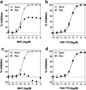
Background: Maraviroc (MVC) is an allosteric CCR5 inhibitor used against HIV-1 infection. While MVC-resistant viruses have been identified in patients, it still remains incompletely known how they adjust their CD4 and CCR5 binding properties to resist MVC inhibition while preserving their replicative capacity. It is thought that they maintain high efficiency of receptor binding. To date however, information about the binding affinities to receptors for inhibitor-resistant HIV-1 remains limited.

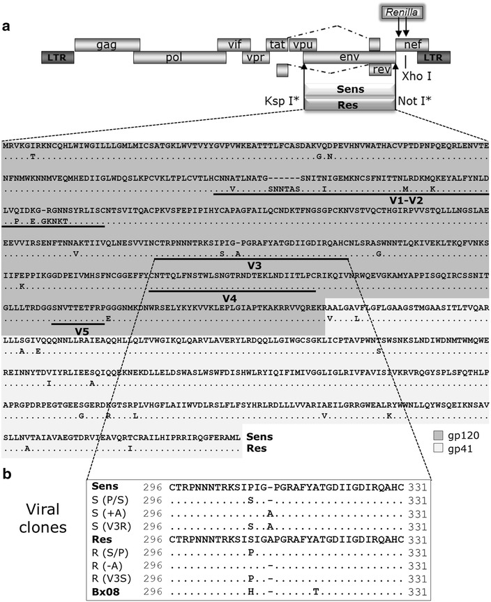
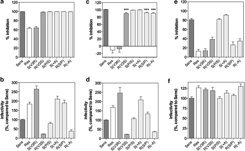
Results: Here, we show by means of viral envelope (gp120) binding experiments and virus-cell fusion kinetics that a MVC-resistant virus (MVC-Res) that had emerged as a dominant viral quasispecies in a patient displays reduced affinities for CD4 and CCR5 either free or bound to MVC, as compared to its MVC-sensitive counterpart isolated before MVC therapy. An alanine insertion within the GPG motif (G310_P311insA) of the MVC-resistant gp120 V3 loop is responsible for the decreased CCR5 binding affinity, while impaired binding to CD4 is due to sequence changes outside V3. Molecular dynamics simulations of gp120 binding to CCR5 further emphasize that the Ala insertion alters the structure of the V3 tip and weakens interaction with CCR5 ECL2. Paradoxically, infection experiments on cells expressing high levels of CCR5 also showed that Ala allows MVC-Res to use CCR5 efficiently, thereby improving viral fusion and replication efficiencies. Actually, although we found that the V3 loop of MVC-Res is required for high levels of MVC resistance, other regions outside V3 are sufficient to confer a moderate level of resistance. These sequence changes outside V3, however, come with a replication cost, which is compensated for by the Ala insertion in V3.

Conclusion: These results indicate that changes in the V3 loop of MVC-resistant viruses can augment the efficiency of CCR5-dependent steps of viral entry other than gp120 binding, thereby compensating for their decreased affinity for entry receptors and improving their fusion and replication efficiencies. This study thus sheds light on unsuspected mechanisms whereby MVC-resistant HIV-1 could emerge and grow in treated patients.


Figures

Figure 1
Figure 1
Cloning, sequence analysis and site-directed mutants of MVC-Sens and MVC-Res Envs. a Schematic representation of the proviral vector pNL-KspI/env/NotI-Ren. The KspI site was introduced in the proviral clone pNL4-3Ren to allow the cloning of MVC-Sens and MVC-Res gp160. Analysis of the MVC-Res Env sequence shows 32 mutations as compared to MVC-Sens Env, 18 within gp120 and 14 within gp41, as well as eight amino acid insertions within gp120. The V3 loop of MVC-Res Env contains two changes, the P308S mutation and an insertion of an Alanine within the GPGR tip (G310_P311insA). The MVC-Sens and MVC-Res Env sequences are similar to those reported in two previous papers, except in the N- and C-terminal parts where we noted several amino acid changes. Indeed, in the sequences used in the references [17] and [33], which are deposited in the Los Alamos HIV Sequence Database, the 41 first residues and the 105 last residues originate from the HxB2 HIV-1 strain. b Amino acid sequences of the V3 loops of the different site-directed mutants of MVC-Sens and MVC-Res used in this study. S and R refer to the parental sequences from which the mutant sequences are derived. Dots indicate residues that are identical to those of the parental Env sequence, and dashes indicate gaps. The sequence of the V3 loop of gp120 from the HIV-1 strain Bx08, to which MVC-Sens and MVC-Res Envs are compared in this study, is also shown. The first Cys residue of the V3 loop is equivalent to C296 in the HXB2 sequence and thus noted as such in the MVC-Sens, MVC-Res and Bx08 V3 sequences.
Figure 2
Figure 2
Susceptibility of MVC-Sens and MVC-Res to inhibition by MVC and TAK 779. U87-CD4/CCR5 cells (a, b) or PBMCs (c, d) were inoculated with equal amounts of MVC-Sens or MVC-Res (10 ng of Gag p24) in the absence or in the presence of increasing concentrations of MVC (a, c) or TAK 779 (b, d). Data points are expressed as percent inhibition of infection relative to control infection measured in the absence of MVC (0%) and were fitted to a sigmoidal dose–response model with a variable slope. Representative experiments out of five independent experiments performed in triplicate are shown.
Figure 3
Figure 3
MVC resistance and viral replicative capacity: The effects of amino acid changes in the MVC-Sens and MVC-Res V3 loops and dependence on the cell type. U87-CD4/CCR5 cells (a, b), PBMCs (c, d) or CD4- and CCR5-expressing HEK 293T cells (e, f) were infected with equal amounts (10 ng of Gag p24) of MVC-Sens, MVC-Res or their related variants S(V3R), R(V3S), S(P/S), S(+A), R(S/P) or R(−A), in the absence or in the presence of 10 μM MVC. The percents of infection inhibition (panels a, c, e) were determined as indicated in the legend of Figure 2. Viral infectivities (b, d, f) were determined by measuring luciferase activity in the cell lysates 30 h (U87, HEK) or 48 h (PBMCs) post-infection and are expressed as percent infectivity relative to that of MVC-Sens (100%). Results are mean ± SEM of 3–8 independent experiments performed in triplicate. ***P < 0.001 in unpaired two-tailed Student t test.
Figure 4
Figure 4
Relative efficiencies of CD4 and CCR5 usage by MVC-Sens and MVC-Res Envs as assessed by the 293-Affinofile receptor affinity profiling system. 293-Affinofile cells were induced by minocycline and/or ponasterone A to express 25 different combinations of CD4 and CCR5 expression levels and then infected by equal amounts (10 ng of Gag p24) of MVC-Sens or MVC-Res in the presence or in the absence of 10 μM MVC. Thirty hours post-infection, luciferase activity was measured in the cell lysates and the three metrics θ (a), M (b) and Δ (c) describing viral infectivity were then determined using the VERSA website (http://versa.biomath.ucla.edu). The maximally induced levels of CD4 and CCR5 were 287,000 and 83,000 receptor/cell. Results represent the mean ± SE of at least four independent determinations. *P < 0.05 in unpaired two-tailed Student t test.
Figure 5
Figure 5
Receptor binding properties of wild-type and modified gp120 monomers derived from the MVC-Sens and MVC-Res viral isolates. a Competition of mAb Q4120 binding to CD4-expressing HEK 293T cells by increasing concentrations of the indicated purified monomeric gp120. Results were normalized for nonspecific binding (0%) and specific binding in the absence of glycoprotein (100%, B0) and were fitted to a one-site competitive binding model. A representative experiment performed in duplicate is shown (n = 3). b Equilibrium saturation binding of the 35S-labeled gp120 of MVC-Sens, MVC-Sens(P/S) (i.e. MVC-Sens wherein Pro-308 is substituted by Ser), MVC-Res(V3S) (i.e. MVC-Res whose V3 loop is replaced by that of MVC-Sens) or MVC-Res(−A) (i.e. MVC-Res lacking the Ala insertion). Curves represent specific binding of glycoproteins to crude membranes from CCR5-expressing HEK 293T cells, determined in the presence of 400 nM sCD4, and obtained by subtracting from total binding the non specific binding measured in the presence of 10 μM TAK779 or using parental HEK cells. Data were fitted to a one-site binding model. Representative experiments performed in duplicate are shown (n = 3–4). c Specific binding of 10 nM of the indicated 35S-labeled gp120 monomers (+400 nM sCD4) to CCR5-expressing HEK cells, in the presence (+) or absence (−) of 10 μM MVC, was calculated as in panel (b), and then expressed as percent of MVC-Sens gp120 binding in the absence of MVC (100%). The binding of glycoproteins measured in the presence of 10 μM TAK779 was considered as nonspecific binding in these experiments. Of note, in some cases, glycoproteins showed levels of binding that came slightly lower than this nonspecific binding, explaining why “negative” specific binding are plotted in the panel. Results represent mean ± SD of 2–5 independent experiments performed in duplicate. d The panel represents specific binding on CD4-expressing HEK cell membranes of the indicated 35S-gp120 used at a concentration equal to their Ki value for CD4 deduced from the displacement experiments of mAb Q4120 binding shown in panel (a) (see text). One experiment out of two is shown. e Binding of the indicated concentrations of MVC-Sens (closed symbols and straight lines) or MVC-Res (open symbols and dashed lines) gp120 to 17b (squares and diamonds) or E51 (circles and triangles) mAbs immobilized on a CM4 sensorchip, alone (triangles and diamonds) or after preincubation with 200 nM sCD4 (circles and squares). Of note, triangles and diamonds are superimposed at the bottom of the panel due to marginal binding in the absence of sCD4. Open and closed squares are also superimposed due to similar binding of MVC-Sens and MVC-Res gp120s to mAb 17b. f and g Competition of 10 nM 35S-gp120Bx08 (f) or 35S-gp120Sens (g) binding to CCR5-expressing membranes by increasing concentrations of the indicated unlabeled gp120 was carried out in the presence of an excess concentration of sCD4 (1,000 nM). Results were normalized for nonspecific binding determined in the presence of 10 μM TAK779 (0%) and specific binding in the absence of competitor (100%, B0) and were fitted to a one-site competitive binding model. Representative experiments out of 2–3 independent experiments run in duplicate are shown.
Figure 6
Figure 6
Characteristics of MVC-Sens and MVC-Res fusion with CD4+ T cells. a Fusion kinetics of BlaM-vpr-containing MVC-Sens and MVC-Res viruses with activated CD4+ T-lymphocytes are shown, in the presence or in the absence of 10 μM MVC. After virus spinoculation onto cells at 4°C and cell washing, fusions were run for the indicated times at 37°C and cells were then loaded with CCF2/AM. Results are expressed as the percentage of BlaM-vpr positive cells, i.e. cells displaying cleaved CCF2/AM fluorescence (at 447 nm). Results are mean ± SEM of two independent determinations out of at least five. b Levels of fusion at 2 h. for the viruses MVC-Sens, MVC-Res and their related variants Sens(+A), Sens(V3R), Res(−A) and Res(V3S), in the absence or in the presence of 10 μM MVC. Results, expressed as fold changes compared to the extent of fusion of MVC-Sens, represent mean ± SEM of 2–10 independent determinations. c The panel represents data from the fusion kinetics of MVC-Sens and MVC-Res w/or w/o 10 μM MVC that were normalized to the maximal extent of fusion at 300 min. d and e Time-of-inhibitor-addition experiments revealing that MVC-Res interacts with CD4 and CCR5 receptors more slowly than MVC-Sens. Fusion of viruses with CD4+ T cells was measured at 240 min under conditions where 50 μg/ml of the anti-CD4 mAb Q4120 (d) or 20 μg/ml of the anti-CCR5 mAb 2D7 (e) was added at the indicated time points after cell transfer to 37°C (time zero), in the presence or in the absence of 10 μM MVC. Results are expressed as the percentage of fusion relative to fusion in the absence of inhibitor (time 240 min). Representative experiments out of three independent experiments are shown.
Figure 7
Figure 7
Organization of the MVC-Sens and MVC-Res V3 tips. The frequency of occurrence of hydrogen bonds in the V3 tips of MVC-Sens (a) and MVC-Res (b) Envs were calculated for every 25 ps segment of each molecular dynamics trajectory. The average rates and standard deviations calculated on the five trajectories are indicated near each H-bond noted with dashed green lines.
Figure 8
Figure 8
Molecular models of CCR5 binding the V3 tip of MVC-Sens Env (left) or the V3 tip of MVC-Res Env and MVC (right). a and b Three-dimensional view of the complexes. The backbone of CCR5 is represented as grey ribbons and the backbone of V3 as green ribbons. The side chains of amino acids involved in inter-molecular interactions are represented as sticks. MVC is represented as sticks. c and d Schematic view of binding modes. Light, medium and dark grey boxes represent the receptor residues in ECL2, N-terminus, and TM7, respectively. H-bonds are indicated with dotted black lines, aromatic stacking with a dotted blue line and ionic bonds with dotted red lines. The thicker the line, the more stable is the interaction.

References

    1. Bachelerie F, Ben-Baruch A, Burkhardt AM, Combadiere C, Farber JM, Graham GJ, et al. International Union of Pharmacology. LXXXIX. Update on the extended family of chemokine receptors and introducing a new nomenclature for atypical chemokine receptors. Pharmacol Rev. 2014;66:1–79. doi: 10.1124/pr.113.007724. - DOI - PMC - PubMed
    1. Cormier EG, Dragic T. The crown and stem of the V3 loop play distinct roles in human immunodeficiency virus type 1 envelope glycoprotein interactions with the CCR5 coreceptor. J Virol. 2002;76:8953–8957. doi: 10.1128/JVI.76.17.8953-8957.2002. - DOI - PMC - PubMed
    1. Huang CC, Tang M, Zhang MY, Majeed S, Montabana E, Stanfield RL, et al. Structure of a V3-containing HIV-1 gp120 core. Science. 2005;310:1025–1028. doi: 10.1126/science.1118398. - DOI - PMC - PubMed
    1. Kwong PD, Wyatt R, Robinson J, Sweet RW, Sodroski J, Hendrickson WA. Structure of an HIV gp120 envelope glycoprotein in complex with the CD4 receptor and a neutralizing human antibody. Nature. 1998;393:648–659. doi: 10.1038/31405. - DOI - PMC - PubMed
    1. Brelot A, Heveker N, Adema K, Hosie MJ, Willett B, Alizon M. Effect of mutations in the second extracellular loop of CXCR4 on its utilization by human and feline immunodeficiency viruses. J Virol. 1999;73:2576–2586. - PMC - PubMed

Publication types