. 2015 Jun 18;16(1):72.
doi: 10.1186/s12931-015-0232-4.

Fibroblast-epithelial cell interactions drive epithelial-mesenchymal transition differently in cells from normal and COPD patients

Affiliations

Fibroblast-epithelial cell interactions drive epithelial-mesenchymal transition differently in cells from normal and COPD patients

Michiyoshi Nishioka et al. Respir Res. .

Abstract

Background: Epithelial-to-mesenchymal transition (EMT), which involves changes in cellular morphology of highly polarized epithelial cells and the gain of mesenchymal cell phenotype with migratory and invasive capacities, is implicated in smoking-related chronic obstructive pulmonary disease (COPD). However, the interactions of fibroblasts and epithelial cells and the participation of fibroblasts in the EMT processes in COPD are poorly understood. Here, we investigated the hypothesis that EMT is active in human bronchial epithelial (HBE) cells of COPD patients, and that mediators secreted by lung fibroblasts from COPD patients induce EMT.

Methods: Primary HBE cells from normal subjects and COPD patients were purchased from LONZA. HLFs were derived from resected lung obtained from normal (N) and COPD (D) subjects and their conditioned medium (CM) was collected after 2-day culture in serum-free medium. The expression of epithelial and mesenchymal markers as well as EMT-related transcription factors in lung biopsies, and in HBE cells following stimulation with CM from both normal human lung fibroblasts (NHLF) and COPD human lung fibroblasts (DHLF) was evaluated by immunohistochemistry, qRT-PCR and western blot.

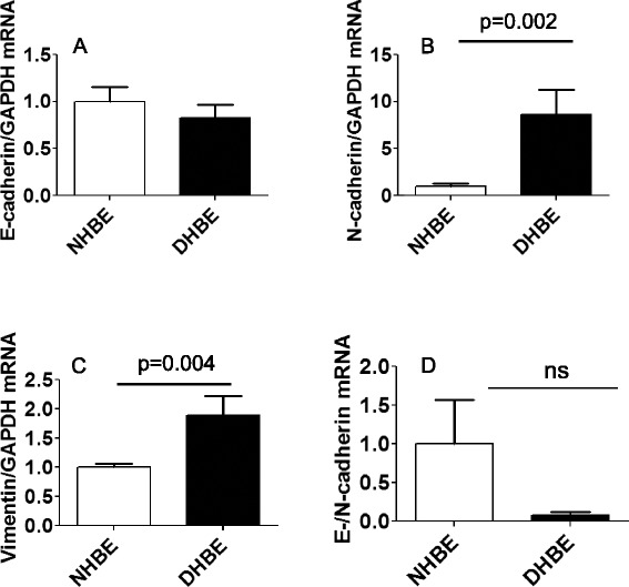
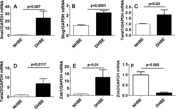
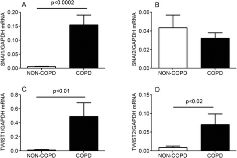
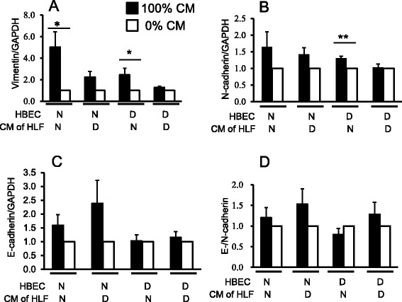
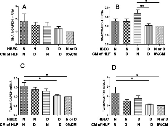
Results: Basal mRNA expression of mesenchymal markers and EMT-related transcription factors were increased in DHBE cells compared to normal human bronchial epithelial cells (NHBE) cells as well as in COPD lungs. CM from NHLF significantly induced vimentin expression in both NHBE and COPD human bronchial epithelial cells (DHBE) cells, but only increased N-cadherin expression in DHBE cells. CM from NHLF significantly induced Twist1 and Twist2 expression in NHBE cells and increased Snai2 (Slug) expression in DHBE cells. While CM from NHLF had no effect on such EMT markers, CM from DHLF significantly increased the protein expression of E-cadherin and vimentin in NHBE cells compared to control. N-cadherin expression was upregulated to a greater degree in NHBE cells than DHBE cells. Only CM from DHLF significantly increased E-/N-cadherin ratio in DHBE cells.

Conclusions: Our results suggest that DHBE cells have partially undergone EMT under baseline conditions. DHLF-CM promoted EMT in NHBE, suggesting that interactions between fibroblast and epithelial cells may play an important role in the EMT process in COPD.


Figures

Fig. 1
Fig. 1
Basal mRNA expression of mesenchymal markers was increased in DHBE cells. Basal mRNA expression of EMT-related markers in non-COPD (NHBE, open bar, n = 3) and COPD (DHBE, closed bar, n = 3) human bronchial epithelial cells were analyzed by real-time RT-PCR. Results were normalized to NHBE and expressed as mean (SE) of three independent experiments. Data were analysed by Student t test, *p < 0.05, compared with NHBE. (a) E-cadherin (b) N-cadherin (c) vimentin (d) Ratio of E-/N-cadherin. ns: not significant
Fig. 2
Fig. 2
Basal mRNA expression of EMT-related transcription factors, except Zeb2, was higher in DHBE cells. Basal mRNA expression of EMT-related transcription factors in non-COPD (NHBE, open bar, n = 3) and COPD (DHBE, closed bar, n = 3) human bronchial epithelial cells were analyzed by real-time RT-PCR. Results were normalized to NHBE and expressed as mean (SE) of three independent experiments. Data were analysed by Student t test, *p < 0.05, **p < 0.01, ***p < 0.0001, compared with NHBE. (a) Snail (b) Slug (c) Twist1 (d) Twist2 (e) Zeb1 and (f) Zeb2
Fig. 3
Fig. 3
mRNA expression analysis of EMT-related transcription factors in non-COPD and COPD lungs. Basal mRNA expression of EMT-related transcription factors in non-COPD (white bars, n = 4) and COPD (black bars, n = 4) subjects. Lung tissues from non-COPD and COPD subjects were analyzed by real-time RT-PCR. Results were normalized to housekeeping gene (GAPDH mRNA) and expressed as mean (SE) of four independent experiments. Data were analysed by unpaired Student’s t test, (a) Snail (b) Snai2 (c) Twist1 (d) Twist2. Non-COPD subjects are non smokers
Fig. 4
Fig. 4
Snail1 and E-cadherin protein expression in non-COPD and COPD lungs. Immunohistochemical localization of EMT markers in the airway epithelium of non-COPD (a, c, e) and COPD (b, d, f) lungs (n = 3). Immunostaining for E-cadherin (a, b) and Snail1 transcription factor (c, d) and N-cadherin (e, f). The dark brown color is indicative of strong expression (denoted by red arrows) of E-cadherin in non-COPD (a), Snail1 in COPD (d) and N-cadherin in COPD (f) lung. Negative control was stained with isotype-matched control antibody (data not shown). Non-COPD subjects are non smokers. aw: airway lumen
Fig. 5
Fig. 5
Basal expression of HGF in non-COPD (NHPF) and COPD (CHPF) lung fibroblasts. mRNA expression (a) and the protein secretion (b) of hepatocyte growth factor in conditioned medium of normal (NHPF) and COPD (CHPF) lung fibroblasts. Results were expressed as mean (SE) of six independent experiments. Data were analysed by unpaired Student’s t test, Non-COPD subjects are non smokers
Fig. 6
Fig. 6
mRNA expression of EMT markers in HBE cells after 3-day treatment with CM from HLF. HBE cells from non-COPD (N) or COPD patients (D) were stimulated with 100 % CM (black bars), or without CM (white bars) collected from human lung fibroblasts (HLF) derived from non-smokers (N) or COPD subjects (D) for 3 days and total RNA was extracted. The mRNA expression of EMT markers were determined by real-time RT-PCR and normalized to GAPDH mRNA expression in the same origin of HBE cells. (a) E-cadherin, (b) N-cadherin, (c) vimentin, and (d) the ratio of E-/N-cadherin were calculated. Data were analysed and expressed as mean (SE) of from six to nine independent experiments by one-way ANOVA with Tukey’s multiple comparison test, *p < 0.05, **p < 0.01, compared between the indicated groups
Fig. 7
Fig. 7
Effects of CM from HLF on protein expression of EMT markers in HBE cells. HBE cells from normal subjects (N) or COPD patients (D) were lysed after stimulation with or without 100 % CM collected from human lung fibroblasts of N or D for 5 days. Total protein was prepared analysed by western blotting, using 40 μg of protein per lane. The membranes were probed with anti-E-cadherin, anti-N-cadherin, anti-vimentin, or anti-GAPDH antibodies. The intensities of the bands for target protein were normalized to the corresponding GAPDH bands for the each treated cells. (a) E-cadherin, (b) N-cadherin, (c) vimentin, and (d) the ratio of E-/N-cadherin were calculated. Data were analysed and expressed as mean (SE) of from seven to ten independent experiments by one-way ANOVA with Tukey’s multiple comparison test, *p < 0.05, **p < 0.01, compared between the indicated groups
Fig. 8
Fig. 8
mRNA expression of EMT-related transcription factors in HBE cells after 3-day treatment with CM from HLF. HBE cells from normal subjects (N) or COPD patients (D) were stimulated with or without 100 % CM collected from human lung fibroblasts of N or D for 3 days and total RNA was extracted. The mRNA expressions of EMT-related transcription factors were determined by real-time RT-PCR and normalized to GAPDH mRNA expression in the same origin of HBE cells. (a) Snail, (b) Slug, (c) Twist1, (d) Twist2, and (e) Zeb1 were calculated. Data were analysed and expressed as mean (SE) of from six to nine independent experiments by one-way ANOVA with Tukey’s multiple comparison test, *p < 0.05, **p < 0.01, compared between the indicated groups

References

    1. Rabe KF, Hurd S, Anzueto A, Barnes PJ, Buist SA, Calverley P, et al. Global strategy for the diagnosis, management, and prevention of chronic obstructive pulmonary disease: GOLD executive summary. Am J Respir Crit Care Med. 2007;176:532–55. doi: 10.1164/rccm.200703-456SO. - DOI - PubMed
    1. Nowrin K, Sohal SS, Peterson G, Patel R, Walters EH. Epithelial-mesenchymal transition as a fundamental underlying pathogenic process in COPD airways: fibrosis, remodeling and cancer. Expert Rev Respir Med. 2014;8:547–59. doi: 10.1586/17476348.2014.948853. - DOI - PubMed
    1. Kalluri R, Neilson EG. Epithelial-mesenchymal transition and its implications for fibrosis. J Clin Invest. 2003;112:1776–84. doi: 10.1172/JCI200320530. - DOI - PMC - PubMed
    1. Sohal SS, Reid D, Soltani A, Ward C, Weston S, Muller HK, et al. Evaluation of epithelial mesenchymal transition in patients with chronic obstructive pulmonary disease. Respir Res. 2011;12:130. doi: 10.1186/1465-9921-12-130. - DOI - PMC - PubMed
    1. Sohal SS, Reid D, Soltani A, Ward C, Weston S, Muller HK, et al. Reticular basement membrane fragmentation and potential epithelial mesenchymal transition is exaggerated in the airways of smokers with chronic obstructive pulmonary disease. Respirology. 2010;15:930–8. doi: 10.1111/j.1440-1843.2010.01808.x. - DOI - PubMed

Publication types

MeSH terms