Review
. 2015 Jun 15;142(12):2094-108.
doi: 10.1242/dev.114215.

Orchestrating liver development

Affiliations
Review

Orchestrating liver development

Miriam Gordillo et al. Development. .

Abstract

The liver is a central regulator of metabolism, and liver failure thus constitutes a major health burden. Understanding how this complex organ develops during embryogenesis will yield insights into how liver regeneration can be promoted and how functional liver replacement tissue can be engineered. Recent studies of animal models have identified key signaling pathways and complex tissue interactions that progressively generate liver progenitor cells, differentiated lineages and functional tissues. In addition, progress in understanding how these cells interact, and how transcriptional and signaling programs precisely coordinate liver development, has begun to elucidate the molecular mechanisms underlying this complexity. Here, we review the lineage relationships, signaling pathways and transcriptional programs that orchestrate hepatogenesis.

Keywords: Cholangiocyte; Hepatoblast; Hepatocyte; Liver development; Transcription factors.


Figures

Fig. 1.
Fig. 1.
Liver structure and cell types. (A) The liver is organized from many lobules, which constitute its functional units. Each lobule is composed of a central vein (CV), from which hepatocyte cords radiate towards portal triads. The portal triad consists of a portal vein, hepatic artery and biliary duct. Hepatocyte cords are single-cell sheets of hepatocytes separated by sinusoids that carry blood from the portal triads to the central vein. (B) Within each lobule are a number of sinusoids, which are discontinuous vessels built from specialized fenestrated endothelial cells of the liver. Stellate (or Ito) cells are located in the space of Disse between the hepatocyte cords and sinusoids. Kupffer cells, which are the specialized macrophages of the liver, also reside in sinusoids. Hepatocytes secrete bile salts into the bile canaliculi that lead to the bile duct. Cholangiocytes are the epithelial cells lining the bile ducts.
Fig. 2.
Fig. 2.
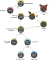
Bipotential progenitors progressively generate hepatic lineages. Four key transition points (A-D) during fetal liver development are highlighted. (A) Bipotential mesendoderm cells segregate into brachyury (BRY)+ mesoderm and definitive endoderm (DE). The prospective foregut endoderm maintains expression of the pioneer transcription factors FOXA2 and GATA4/6 (denoted GATA). (B) Studies of ESC cultures suggest that the DE includes a subset of KDR+ progenitors that generate and support the development of hepatic cells; they can also give rise to CD31 (PECAM1)+ endothelial cells. (C) Foregut DE generates a bipotential hepato-pancreatic progenitor (SOX17+, HHEX+, GATA+) that produces both PDX1+ pancreatic progenitors and HNF1β+, HNF4α+, PDX1 hepatoblasts. It is not known how closely the hepatic cells derived from KDR+ endoderm are related to this cell (question mark). (D) Finally, the hepatoblast is a bipotential progenitor for both cholangiocytes (bile duct cells; HNF6+, SOX9+, HNF1β+) and hepatocytes (PROX1+, HNF4α+). Note that the figure only indicates when factors initially function for transitions; in many cases (such as for FoxA and GATA factors) they continue to be expressed and also function at later stages.
Fig. 3.
Fig. 3.
Liver diverticulum and bud formation in mouse. (A) Sagittal section of the cephalic portion of the E8.25 mouse prospective hepatic endoderm (HE, green). The convergence of the cardiac mesoderm (blue) and septum transversum (ST, purple) is required for hepatic specification. (B-D) Transverse sections of the mouse liver diverticulum progressing to the liver bud stage. (B) At E8.75, endothelial cells (ECs, orange) are found surrounding the thickened hepatic endoderm, which initiates a budding process into the septum transversum. Endothelial cells contribute to hepatic specification. (C) At E9, the hepatic endoderm transitions from a columnar to a pseudostratified epithelium. (D) At E10, hepatic endoderm cells, identified as hepatoblasts, proliferate and migrate into the septum transversum to form the liver bud. Endothelial cells are also required for liver bud formation. Hematopoietic progenitor cells now start to migrate into the bud to establish liver fetal hematopoiesis.
Fig. 4.
Fig. 4.
Bile duct development. Prior to bile duct development, hepatoblasts are the only epithelial progenitor cells in the fetal liver. Ductal plate formation is the first sign of bile duct development. It is initiated at E13.5 in mice and at around day 56-58 after fertilization in humans. The ductal plate is composed of a monolayer of cholangiocyte precursors derived from hepatoblasts in contact with the portal mesenchyme. These precursors express high levels of CK19 compared with hepatoblasts that are located further away from the portal veins. At ∼E17.5 in mice, and during the second trimester in humans, the ductal plate duplicates. Luminal pockets develop between the two layers of precursors. In the perinatal period in mice and at ∼30 weeks of gestation in humans, some of these pockets form bile ducts composed of mature cholangiocytes that maintain CK19 expression. The remaining ductal plate areas regress in a process called ductal plate remodeling. The hepatoblasts located away from the portal veins differentiate into mature hepatocytes that completely downregulate CK19.
Fig. 5.
Fig. 5.
Key signals mediating progenitor fate decisions during hepatogenesis. (A) Activation of Nodal signaling in the embryonic epiblast (Ep) lineage initiates mesendoderm (ME) formation. In Xenopus and zebrafish, maternal signals activate this pathway. In mice, Nodal, BMP4 and WNT3 act in a reinforcing loop to activate Nodal and induce ME. Subsequently, high levels of Nodal signaling establish the endoderm regulatory network leading to the segregation of, and commitment to, definitive endoderm (DE), while FGF and BMP drive mesoderm (M) formation by antagonizing Nodal signaling. (B) The graded activity of Wnt, FGF and BMP signaling patterns the endoderm along the AP axis to generate posterior foregut precursors (PFG) with hepato-pancreatic potential that can be distinguished from anterior foregut [AFG; deriving lung (L) and thyroid (T)] and midgut-hindgut [MG-HG; deriving intestine (I)] progenitors. P represents pancreatic progenitors. (C) FGF-MAPK, BMP and Wnt signaling positively regulate hepatic specification to generate hepatoblasts (Hb), although the role of Wnt at this stage of liver development has not been demonstrated in mammals (indicated by dashed box). (D) Hepatoblast differentiation into cholangiocytes (Ch) and hepatocytes (H), and the final maturation of these cells, is regulated by a wide array of signaling pathways that display complex cross-regulation. These pathways also influence the 3D structural organization of the liver and define its zonal characteristics. The corresponding developmental stages are indicated beneath the scheme.

References

    1. Alder O., Cullum R., Lee S., Kan A. C., Wei W., Yi Y., Garside V. C., Bilenky M., Griffith M., Morrissy A. S. et al. (2014). Hippo signaling influences HNF4A and FOXA2 enhancer switching during hepatocyte differentiation. Cell Rep. 9, 261-271. 10.1016/j.celrep.2014.08.046 - DOI - PMC - PubMed
    1. Ang S.-L. and Rossant J. (1994). HNF-3 beta is essential for node and notochord formation in mouse development. Cell 78, 561-574. 10.1016/0092-8674(94)90522-3 - DOI - PubMed
    1. Angelo J. R., Guerrero-Zayas M.-I. and Tremblay K. D. (2012). A fate map of the murine pancreas buds reveals a multipotent ventral foregut organ progenitor. PLoS ONE 7, e40707 10.1371/journal.pone.0040707 - DOI - PMC - PubMed
    1. Antoniou A., Raynaud P., Cordi S., Zong Y., Tronche F., Stanger B. Z., Jacquemin P., Pierreux C. E., Clotman F. and Lemaigre F. P. (2009). Intrahepatic bile ducts develop according to a new mode of tubulogenesis regulated by the transcription factor SOX9. Gastroenterology 136, 2325-2333. 10.1053/j.gastro.2009.02.051 - DOI - PMC - PubMed
    1. Ben-Haim N., Lu C., Guzman-Ayala M., Pescatore L., Mesnard D., Bischofberger M., Naef F., Robertson E. J. and Constam D. B. (2006). The nodal precursor acting via activin receptors induces mesoderm by maintaining a source of its convertases and BMP4. Dev. Cell 11, 313-323. 10.1016/j.devcel.2006.07.005 - DOI - PubMed

Publication types