Clinical Trial
. 2015 Oct;35(10):1570-8.
doi: 10.1038/jcbfm.2015.125. Epub 2015 Jun 17.

Hippocampal structure, metabolism, and inflammatory response after a 6-week intense aerobic exercise in healthy young adults: a controlled trial

Affiliations
Clinical Trial

Hippocampal structure, metabolism, and inflammatory response after a 6-week intense aerobic exercise in healthy young adults: a controlled trial

Gerd Wagner et al. J Cereb Blood Flow Metab. 2015 Oct.

Abstract

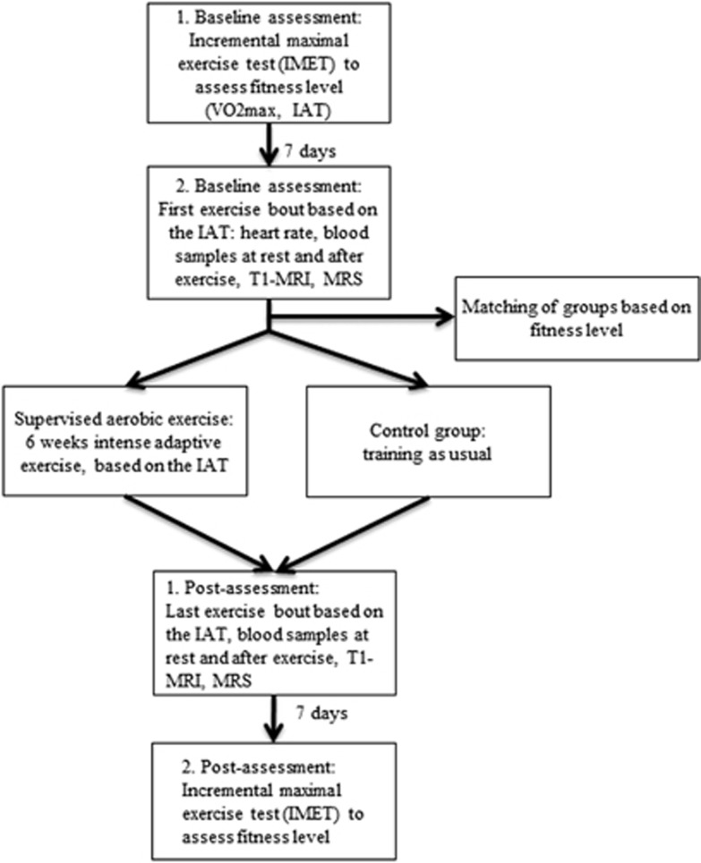
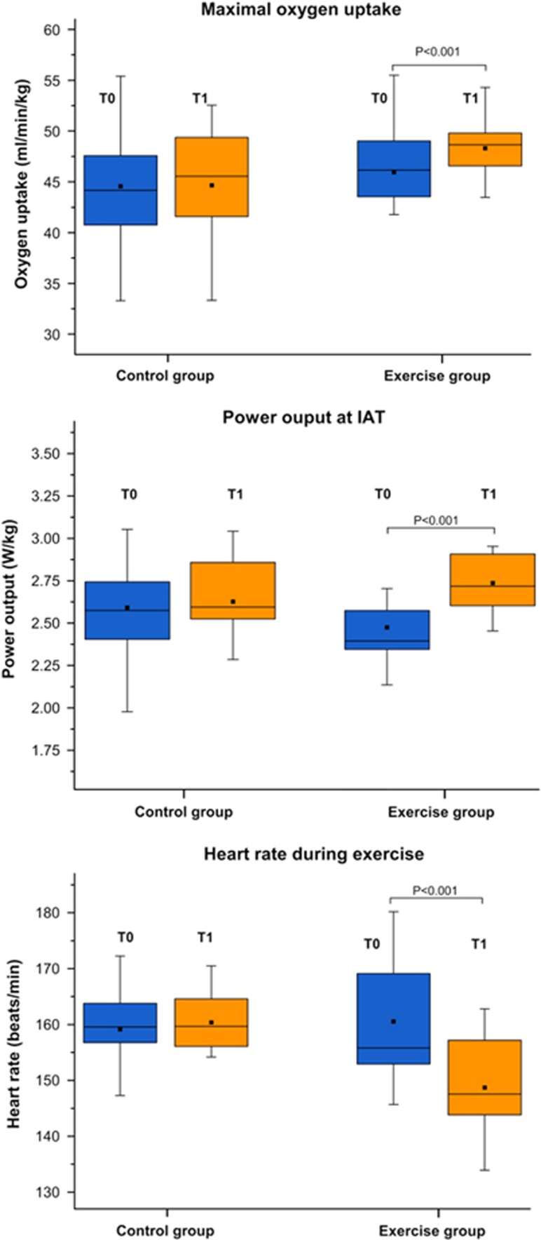
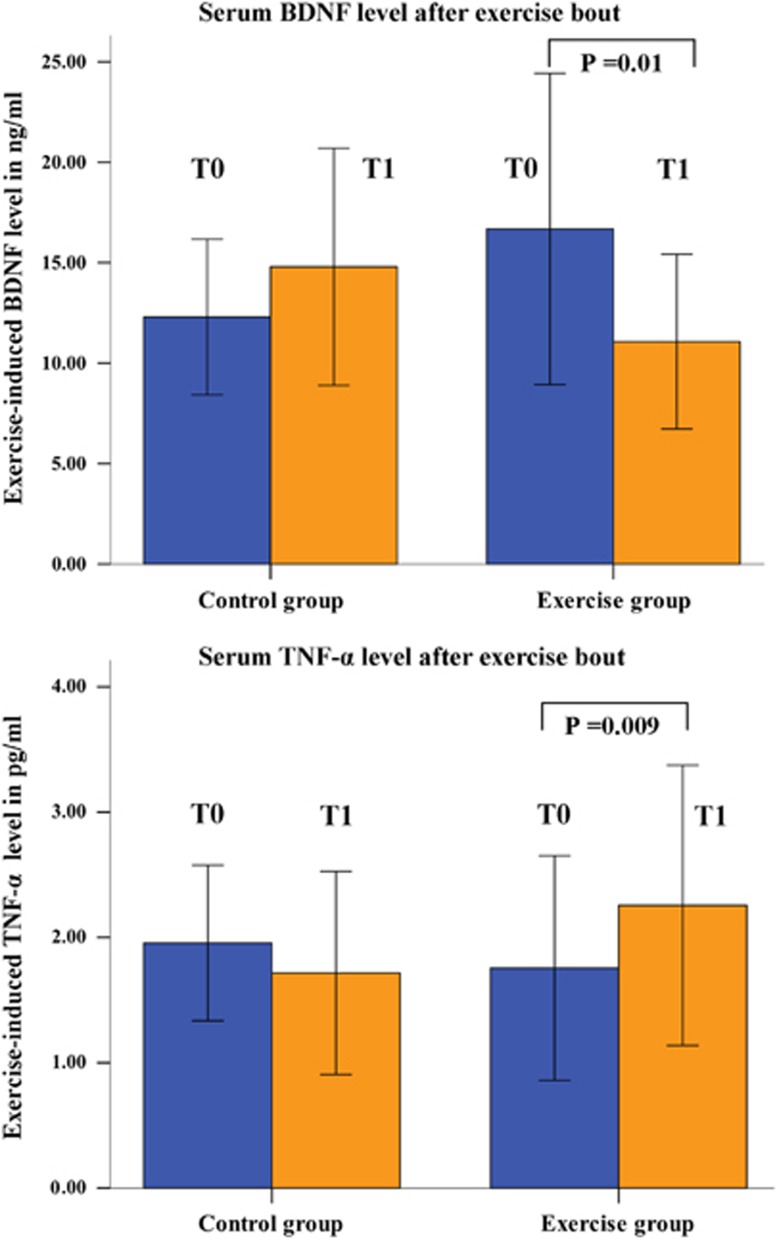
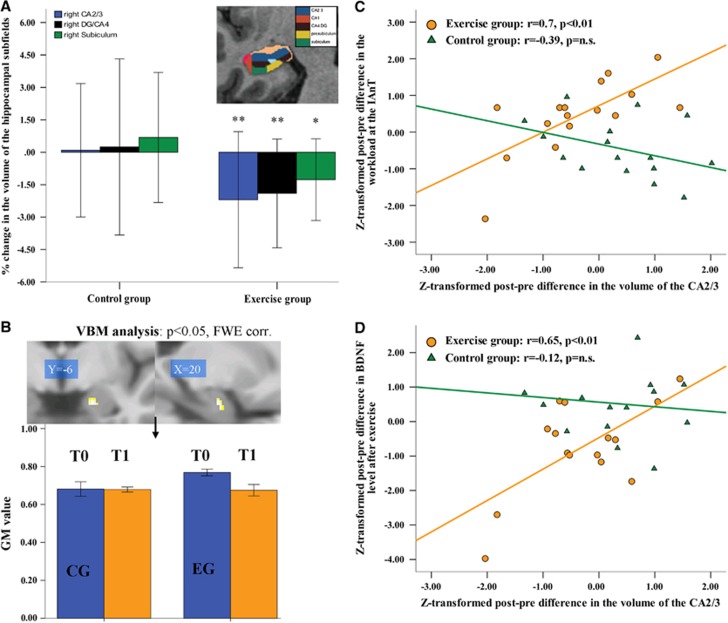
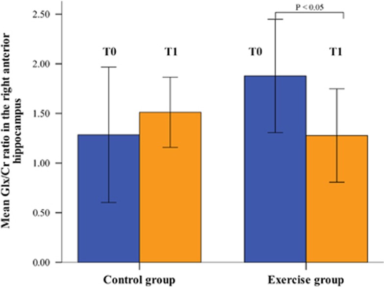
Interventional studies suggest that changes in physical fitness affect brain function and structure. We studied the influence of high intensity physical exercise on hippocampal volume and metabolism in 17 young healthy male adults during a 6-week exercise program compared with matched controls. We further aimed to relate these changes to hypothesized changes in exercised-induced brain-derived neurotrophic factor (BDNF), interleukin-6 (IL-6), and tumor necrosis factor alpha (TNF-α). We show profound improvement of physical fitness in most subjects and a positive correlation between the degree of fitness improvement and increased BDNF levels. We unexpectedly observed an average volume decrease of about 2%, which was restricted to right hippocampal subfields CA2/3, subiculum, and dentate gyrus and which correlated with fitness improvement and increased BDNF levels negatively. This result indicates that mainly those subjects who did not benefit from the exercise program show decreased hippocampal volume, reduced BDNF levels, and increased TNF-α concentrations. While spectroscopy results do not indicate any neuronal loss (unchanged N-acetylaspartate levels) decreased glutamate-glutamine levels were observed in the right anterior hippocampus in the exercise group only. Responder characteristics need to be studied in more detail. Our results point to an important role of the inflammatory response after exercise on changes in hippocampal structure.


Figures

Figure 1
Figure 1
Flow chart of the study. IAT, individual anaerobic threshold; MRI, magnetic resonance imaging; MRS, magnetic resonance spectroscopy.
Figure 2
Figure 2
The box plots illustrate pre-post changes in the maximal oxygen uptake (VO2max), in the power output (W/kg) at the individual anaerobic threshold (IAT) and in heart rate during exercise.
Figure 3
Figure 3
Change in the exercise-induced (as measured immediately after exercise bout) serum brain-derived neurotrophic factor (BDNF) and tumor necrosis factor alpha (TNF-α) concentration (T0 before intervention; T1 after intervention).
Figure 4
Figure 4
(A) Changes in the volumes of the hippocampal subfields CA2/3, dentate gyrus (DG/CA4), and subiculum on the right side as determined by Freesurfer longitudinal data processing (*p≤0.05, **p≤0.01); (B) a significant GROUP × TIME interaction (post-hoc t-test) in the right anterior hippocampus (family-wise error corrected at the voxel and cluster levels, masked for the right hippocampus) as revealed by voxel-based morphometry (VBM). The bar graph illustrates changes in gray matter (GM) volume (T0 before intervention; T1 after intervention); (C) correlations analysis between Z-transformed post-pre differences in the CA2/3 volume and Z-transformed post-pre differences in the power output at the individual anaerobic threshold (exercise group=orange dots, control group (CG)=green triangles); (D) correlation analysis between Z-transformed post-pre differences in the CA2/3 volume and Z-transformed post-pre differences in the BDNF serum concentration as measured immediately after exercise bout (in the exercise group=orange dots; CG=green triangles).
Figure 5
Figure 5
Change in the glutamate+glutamine to creatine (Glx:Cr) ratio measured with magnetic resonance spectroscopy in the right anterior hippocampus (T0 before intervention; T1 after intervention).

References

    1. 1van Praag H, Shubert T, Zhao C, Gage FH. Exercise enhances learning and hippocampal neurogenesis in aged mice. J Neurosci 2005; 25: 8680–8685. - PMC - PubMed
    1. 2Pereira AC, Huddleston DE, Brickman AM, Sosunov AA, Hen R, McKhann GM et al. An in vivo correlate of exercise-induced neurogenesis in the adult dentate gyrus. Proc Natl Acad Sci USA 2007; 104: 5638–5643. - PMC - PubMed
    1. 3Chaddock L, Erickson KI, Prakash RS, Kim JS, Voss MW, Vanpatter M et al. A neuroimaging investigation of the association between aerobic fitness, hippocampal volume, and memory performance in preadolescent children. Brain Res 2010; 1358: 172–183. - PMC - PubMed
    1. 4Erickson KI, Prakash RS, Voss MW, Chaddock L, Hu L, Morris KS et al. Aerobic fitness is associated with hippocampal volume in elderly humans. Hippocampus 2009; 19: 1030–1039. - PMC - PubMed
    1. 5Erickson KI, Voss MW, Prakash RS, Basak C, Szabo A, Chaddock L et al. Exercise training increases size of hippocampus and improves memory. Proc Natl Acad Sci USA 2011; 108: 3017–3022. - PMC - PubMed

Publication types

MeSH terms