Review
. 2015 Jun 2:6:545.
doi: 10.3389/fmicb.2015.00545. eCollection 2015.

The orisome: structure and function

Affiliations
Review

The orisome: structure and function

Alan C Leonard et al. Front Microbiol. .

Abstract

During the cell division cycle of all bacteria, DNA-protein complexes termed orisomes trigger the onset of chromosome duplication. Orisome assembly is both staged and stringently regulated to ensure that DNA synthesis begins at a precise time and only once at each origin per cycle. Orisomes comprise multiple copies of the initiator protein DnaA, which oligomerizes after interacting with specifically positioned recognition sites in the unique chromosomal replication origin, oriC. Since DnaA is highly conserved, it is logical to expect that all bacterial orisomes will share fundamental attributes. Indeed, although mechanistic details remain to be determined, all bacterial orisomes are capable of unwinding oriC DNA and assisting with loading of DNA helicase onto the single-strands. However, comparative analysis of oriCs reveals that the arrangement and number of DnaA recognition sites is surprisingly variable among bacterial types, suggesting there are many paths to produce functional orisome complexes. Fundamental questions exist about why these different paths exist and which features of orisomes must be shared among diverse bacterial types. In this review we present the current understanding of orisome assembly and function in Escherichia coli and compare the replication origins among the related members of the Gammaproteobacteria. From this information we propose that the diversity in orisome assembly reflects both the requirement to regulate the conformation of origin DNA as well as to provide an appropriate cell cycle timing mechanism that reflects the lifestyle of the bacteria. We suggest that identification of shared steps in orisome assembly may reveal particularly good targets for new antibiotics.

Keywords: DNA binding proteins; DNA replication; DnaA; oriC; orisomes; pre-replication complexes; replication origin.


Figures
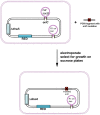
FIGURE 1
FIGURE 1
Scheme of the oriC-specific recombineering method. Recombineering strains harbor sacB and cat genes replacing all of the oriC sequence, inducible genes encoding the lambda RED system, and a plasmid origin of replication (R1ori) linked to a kanamycin resistance determinant. The strain also has a deletion in the dnaA gene. For insertion of oriC mutants into the chromosome, a PCR fragment carrying the desired mutation is electroporated to transform cells in which the RED system was induced. Recombination results in replacement of the sacB and cat genes with the mutated oriC. Successful recombineering confers the ability to grow in the presence of sucrose, and sensitivity to chloramphenicol.
FIGURE 2
FIGURE 2
Map of Escherichia coli oriC and conformation of bORC. (A) The oriC region is mapped, showing positions of binding sites for DnaA, IHF, and Fis, as well as the right (R), middle (M), and left (L) 13mer sequences in the DNA unwinding element (DUE). The three high-affinity sites R1, R2, and R4 are designated by royal blue squares, and the low-affinity sites are marked by small light blue or red rectangles. The red rectangles designate sites that preferentially bind DnaA–ATP, while the sites marked by light blue rectangles bind both nucleotide forms of DnaA equivalently. Small arrowheads show orientation of sites, the two between sites indicates the number of bp separating the sites. Arrows under the map indicate growth direction of DnaA oligomers. The dotted line marks that the oligomer does not span the region between R1 and R5M. Two genes, gidA and mioC, flanking oriC are shown, with the green arrows marking the direction of transcription. (B) Proposed looped conformation of bORC in E. coli. OriC DNA (ribbon) is constrained by DnaA (gray-blue figures in center of loop) bound at R1, R2, and R4 sites, as labeled. Interaction among the three bound DnaAs proposed to be via domain I. The green triangle represents Fis bound to its cognate site.
FIGURE 3
FIGURE 3
Model of staged orisome assembly. Stage 1 (bORC): after initiation of chromosome replication, DnaA rebinds to high affinity R1, R2, and R4 sites. Fis is also bound at this stage, but IHF is not. Low affinity sites are unoccupied. Stage 2: DnaA bound to R4 recruits DnaA for binding to C1. DnaA then progressively fills the remaining arrayed sites, forming an oligomer in the gap region between R2 and R4. The DnaA oligomer displaces Fis, and loss of Fis allows IHF to bind to its cognate site. Stage 3: the bend induced by IHF binding allows DnaA, recruited by R1, to bind to R5M, and form a cross-strand DnaA interaction. A DnaA oligomer then progressively grows toward R2, bound to arrayed low affinity sites, and anchored by R2. In this configuration, oriC DNA is unwound in the DUE, and DnaA in the form of a compact filament binds to the ssDNA.
FIGURE 4
FIGURE 4
Comparison of the number and placement of high affinity DnaA binding sites in oriCs in related members of the Gammaproteobacteria family. The high affinity sites in the oriC regions of E. coli and several related bacterial types are shown. Blue rectangles indicate sites that match the consensus 5′-TTATCCACA, and the pink rectangles mark sites which deviate from this sequence at one or two bases. The arrowheads mark the presumptive orientation of the sites, and the numbers designate the number of base pairs in the gap regions between sites. The brackets below the maps show an E. coli-like arrangement of high affinity sites (see text for details). The two bacterial types below the double line are larger than those above, but it should be noted that the maps are not drawn to scale.

References

    1. Bell S. P. (2002). The origin recognition complex: from simple origins to complex functions. Genes Dev. 16 659–672. 10.1101/gad.969602 - DOI - PubMed
    1. Bonilla C. Y., Grossman A. D. (2012). The primosomal protein DnaD inhibits cooperative DNA binding by the replication initiator DnaA in Bacillus subtilis. J. Bacteriol. 194 5110–5117. 10.1128/JB.00958-12 - DOI - PMC - PubMed
    1. Boonstra M., de Jong I. G., Scholefield G., Murray H., Kuipers O. P., Veening J. W. (2013). Spo0A regulates chromosome copy number during sporulation by directly binding to the origin of replication in Bacillus subtilis. Mol. Microbiol. 87 925–938. 10.1111/mmi.12141 - DOI - PubMed
    1. Bramhill D., Kornberg A. (1988). Duplex opening by dnaA protein at novel sequences in initiation of replication at the origin of the E. coli chromosome. Cell 52 743–755. 10.1016/0092-8674(88)90412-6 - DOI - PubMed
    1. Campbell J. L., Kleckner N. (1990). E. coli oriC and the dnaA gene promoter are sequestered from dam methyltransferase following the passage of the chromosomal replication fork. Cell 62 967–979. 10.1016/0092-8674(90)90271-F - DOI - PubMed

LinkOut - more resources