Review
. 2015 Jun 2:6:553.
doi: 10.3389/fmicb.2015.00553. eCollection 2015.

Nucleocytoplasmic transport of nucleocapsid proteins of enveloped RNA viruses

Affiliations
Review

Nucleocytoplasmic transport of nucleocapsid proteins of enveloped RNA viruses

Wahyu N Wulan et al. Front Microbiol. .

Abstract

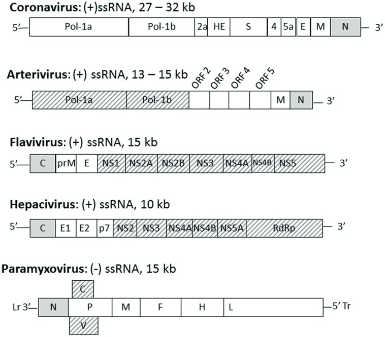
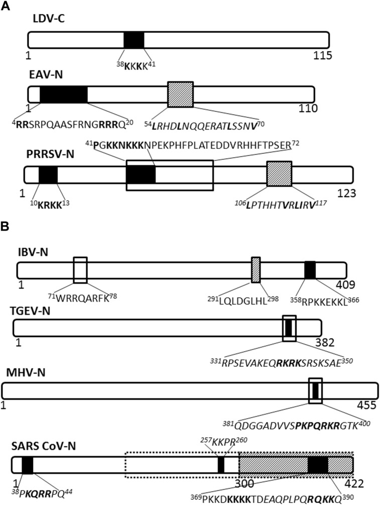
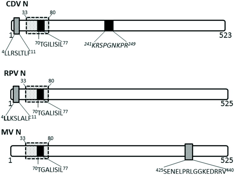
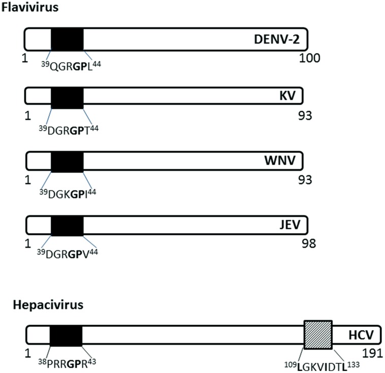
Most viruses with non-segmented single stranded RNA genomes complete their life cycle in the cytoplasm of infected cells. However, despite undergoing replication in the cytoplasm, the structural proteins of some of these RNA viruses localize to the nucleus at specific times in the virus life cycle, primarily early in infection. Limited evidence suggests that this enhances successful viral replication by interfering with or inhibiting the host antiviral response. Nucleocapsid proteins of RNA viruses have a well-established, essential cytoplasmic role in virus replication and assembly. Intriguingly, nucleocapsid proteins of some RNA viruses also localize to the nucleus/nucleolus of infected cells. Their nuclear function is less well understood although significant advances have been made in recent years. This review will focus on the nucleocapsid protein of cytoplasmic enveloped RNA viruses, including their localization to the nucleus/nucleolus and function therein. A greater understanding of the nuclear localization of nucleocapsid proteins has the potential to enhance therapeutic strategies as it can be a target for the development of live-attenuated vaccines or antiviral drugs.

Keywords: antiviral responses; enveloped RNA viruses; nuclear localization; nucleocapsid protein; nucleolar localization.


Figures

FIGURE 1
FIGURE 1
The position of nucleocapsid protein in the genome of selected enveloped RNA virus families. Genomes of the named virus families are indicated schematically with each gene indicated by a box with family name, genome type and length indicated above each schematic. The gene for nucleocapsid protein (N or C) is indicated by a gray box, other structural proteins are indicated by white boxes and non-structural proteins are indicated by hashed boxes. Lr, leader; Tr, trailer.
FIGURE 2
FIGURE 2
Nuclear/nucleolar transport signals in nucleocapsid proteins of Arteriviridae and Coronaviridae. The nucleocapsid proteins of representatives of named virus families are shown schematically. Numbers under the schematics indicate position of amino acid residues, nuclear localization signals (NLSs) are indicated by solid black filled boxes, nuclear export signals (NESs) are indicated by hatched boxes and nucleolus localization/retention signals (NoLS/RS) are indicated by black open boxes. The sequence of the transport signals is shown in single letter format, with essential residues in bold; sequences in italics are predicted but not yet confirmed. (A) Arteriviridae: LDV, lactate dehydrogenase virus; EAV, equine arteritis virus; PRRSV, porcine respiratory and reproductive syndrome virus. (B) Coronaviridae: Broken black line indicates predicted NoLS/RS. IBV, infectious bronchitis virus; TGEV, transmissible gastroenteritis virus; MHV, mouse hepatitis virus; SARS-CoV, severe acute respiratory syndrome coronavirus.
FIGURE 3
FIGURE 3
Nuclear transport signals in nucleocapsid proteins of Paramyxoviridae. The NLSs and NESs are indicated as in Figure 2; a dashed-line box with dots indicates flanking sequences that have been shown to have a role in nuclear import and retention. CDV, canine distemper virus; RPV, Rinderpest virus; MV, measles virus.
FIGURE 4
FIGURE 4
Nuclear localization signals in C (capsid or core) proteins of Flaviviridae. The NLSs for each viral protein and the NES for HCV are indicated as in Figure 2. DENV-2, Dengue virus serotype 2, KV, Kunjin virus; WNV, West Nile virus; JEV, Japanese encephalitis virus; HCV, hepatitis C virus.

References

    1. Ahmed M., Lyles D. S. (1998). Effect of vesicular stomatitis virus matrix protein on transcription directed by host RNA polymerases I, II, and III. J. Virol. 72 8413–8419. - PMC - PubMed
    1. Ahmed M., Mckenzie M. O., Puckett S., Hojnacki M., Poliquin L., Lyles D. S. (2003). Ability of the matrix protein of vesicular stomatitis virus to suppress beta interferon gene expression is genetically correlated with the inhibition of host RNA and protein synthesis. J. Virol. 77 4646–4657. 10.1128/JVI.77.8.4646-4657.2003 - DOI - PMC - PubMed
    1. Andersen J. S., Lyon C. E., Fox A. H., Leung A. K., Lam Y. W., Steen H., et al. (2002). Directed proteomic analysis of the human nucleolus. Curr. Biol. 12 1–11. 10.1016/S0960-9822(01)00650-9 - DOI - PubMed
    1. Azmi A. S., Aboukameel A., Bao B., Sarkar F. H., Philip P. A., Kauffman M., et al. (2013a). Selective inhibitors of nuclear export block pancreatic cancer cell proliferation and reduce tumor growth in mice. Gastroenterology 144 447–456. 10.1053/j.gastro.2012.10.036 - DOI - PMC - PubMed
    1. Azmi A. S., Al-Katib A., Aboukameel A., Mccauley D., Kauffman M., Shacham S., et al. (2013b). Selective inhibitors of nuclear export for the treatment of non-Hodgkin’s lymphomas. Haematologica 98 1098–1106. 10.3324/haematol.2012.074781 - DOI - PMC - PubMed