Review
. 2015 May 29:4:212281.
doi: 10.7573/dic.212281. eCollection 2015.

Diagnosis and management of lentigo maligna: a review

Affiliations
Review

Diagnosis and management of lentigo maligna: a review

Julia M Kasprzak et al. Drugs Context. .

Abstract

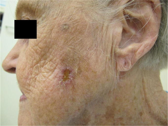
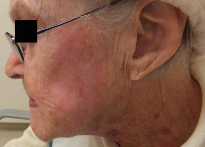
Lentigo maligna is a melanocytic neoplasm occurring on sun-exposed skin, usually on the head and neck, of middle-aged and elderly patients. It is thought to represent the in situ phase of lentigo maligna melanoma. The ill-defined nature and potentially large size of lesions can pose significant diagnostic and treatment challenges. The goal of therapy is to cure the lesions in order to prevent development of invasive disease, and surgical excision is the treatment of choice to achieve clear histological margins. Nonsurgical treatment modalities have been reported; however, evidence is lacking to support their use. Age, general health, and comorbidities need to be taken into account when deciding the right treatment modality for each individual patient.

Keywords: Mohs micrographic surgery; diagnosis; histopathology; imiquimod; immunohistochemistry; lentigo maligna; staged excision.


Figures

Figure 1A.
Figure 1A.
Lentigo maligna was diagnosed with a 4 mm punch biopsy on the left cheek. Close examination under the Wood’s lamp showed that the small biopsy was within a large brown patch with color variegation and ill-defined margin, and multiple scattered brown macules.
Figure 1B.
Figure 1B.
Multiple scouting biopsies were taken from the pigmented patch and macules for histological evaluation. Among twelve sampled areas, five were lentigo maligna, one was atypical junctional melanocytic proliferation, and the rest were pigmented actinic keratoses.
Figure 1C.
Figure 1C.
Design for the first stage slow Mohs excision. A 3 mm margin debulking excision was taken down to subcutaneous fat for evaluation of Breslow depth via vertical sectioning, and another 3 mm margin was taken down to deep subcutaneous fat or fascia for complete margin evaluation via horizontal en-face sectioning.
Figure 1D.
Figure 1D.
Defect after the first stage slow Mohs excision.
Figure 1E.
Figure 1E.
Partial simple closure after the first stage slow Mohs excision before the patient was discharged home.
Figure 1F.
Figure 1F.
A narrow strip of control skin was taken from the contralateral, normal appearing, sun-damaged area and submitted for en-face sectioning.
Figure 1G.
Figure 1G.
Defect after the second stage slow Mohs excision. Noted that additional peripheral margin was taken around the tumor involved margin by the temple hair line.
Figure 1H.
Figure 1H.
Three weeks post a large cervical facial flap reconstruction (courtesy of Dr. Bradley Manning).
Figure 2A.
Figure 2A.
Hematoxylin and eosin stain of the original biopsy of an eyelid lentigo maligna demonstrated classic lentigo maligna or melanoma in situ. There are an increased number of melanocytes at the dermoepidermal junction forming nests, as well as presenting as single cells with mildly enlarged, hyperchromatic nuclei (10X).
Figure 2B.
Figure 2B.
The higher power of 2A (20X).
Figure 2C.
Figure 2C.
An example of positive histological margin of lentigo maligna following slow Mohs excision. Immunohistochemistry stains using MART-1 (and MiTF, another melanocytic marker; photos not shown) strongly highlight an increased number of melanocytes at the dermoepidermal junction. There is multifocal confluence and there are scattered small nests. There is also prominent extension along adnexal epithelium.
Figure 2D.
Figure 2D.
An example of histological morphology of normal appearing, chronically sun-exposed skin taken from the patient’s contralateral cheek. In this control skin, there is also a moderately increased number of melanocytes, some of which confluent, but there are no nests or pagetoid spreads.
Figure 3.
Figure 3.
Schematic illustration of bread loaf sections for evaluating tissue margins. Vertical sections 1, 2, 3 are taken from the middle and at both ends of tissue blocks (A, B, C, and D). All three vertical sections appear clear of tumor; however, there is residual cancer in block B that is not included and therefore missed in representative vertical sections, creating a false negative margin (reprinted with permission from Dr. Stephen Snow, Mohs Micrographic Surgery, 2nd Edition, The University of Wisconsin Press, 2004).
Figure 4.
Figure 4.
Schematic illustration of horizontal en-face sections of tissue during Mohs micrographic surgery. Tissue blocks A and B were processed horizontally from deeper portion of the specimen toward the superficial portion representing epidermis. Each horizontal sectioning includes the deep and lateral edges for a complete margin evaluation. Cancer is found toward the center of the tissue block B, which is documented with color-coding in a map for orientation of each block (reprinted with permission from Dr. Stephen Snow, Mohs Micrographic Surgery, 2nd Edition, The University of Wisconsin Press, 2004).
Figure 5A.
Figure 5A.
Subtotal shave biopsy of an ill-defined brown patch on the left cheek confirmed the diagnosis of lentigo maligna in this elderly woman.
Figure 5B.
Figure 5B.
The patient was treated with imiquimod 5% cream five times weekly for approximately two months. At her three-month post-treatment follow-up, she had mild hypopigmentation and erythema on the left cheek.

Similar articles

Cited by

References

    1. van Ruth S, Toonstra J. Eponyms of Sir Jonathan Hutchinson. Int J Dermatol. 2008;47(7):754–8. http://dx.doi.org/10.1111/j.1365-4632.2008.03696.x. - DOI - PubMed
    1. Gaudy-Marqueste C, Madjlessi N, Guillot B, Avril MF, Grob JJ. Risk factors in elderly people for lentigo maligna compared with other melanomas: a double case-control study. Arch Dermatol. 2009;145(4):418–23. http://dx.doi.org/10.1001/archdermatol.2009.1. - DOI - PubMed
    1. Purdue MP, From L, Kahn HJ, Armstrong BK, Kricker A, Gallagher RP, McLaughlin JR, Klar NS, Marrett LD. Etiologic factors associated with p53 immunostaining in cutaneousmalignant melanoma. Int J Cancer. 2005;117(3):486–93. http://dx.doi.org/10.1002/ijc.21196. - DOI - PubMed
    1. Sasaki Y, Niu C, Makino R, Kudo C, Sun C, Watanabe H, Matsunaga J, Takahashi K, Tagami H, Aiba S, Horii A. BRAF point mutations in primary melanoma show different prevalences by subtype. J Invest Dermatol. 2004;123(1):177–83. http://dx.doi.org/10.1111/j.0022-202X.2004.22722.x. - DOI - PubMed
    1. Maldonado JL, Fridlyand J, Patel H, Jain AN, Busam K, Kageshita T, Ono T, Albertson DG, Pinkel D, Bastian BC. Determinants of BRAF mutations in primary melanomas. J Natl Cancer Inst. 2003;95(24):1878–90. - PubMed

LinkOut - more resources