Review
. 2015 Jun 2:5:48.
doi: 10.3389/fcimb.2015.00048. eCollection 2015.

Purification and proteomics of pathogen-modified vacuoles and membranes

Affiliations
Review

Purification and proteomics of pathogen-modified vacuoles and membranes

Jo-Ana Herweg et al. Front Cell Infect Microbiol. .

Abstract

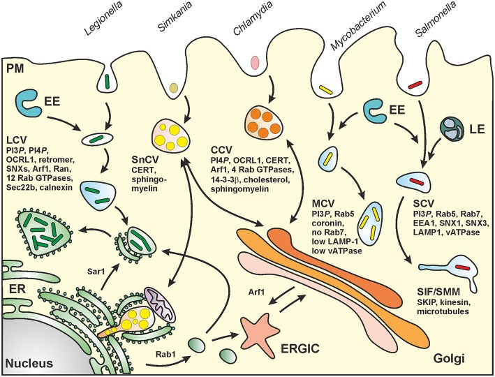
Certain pathogenic bacteria adopt an intracellular lifestyle and proliferate in eukaryotic host cells. The intracellular niche protects the bacteria from cellular and humoral components of the mammalian immune system, and at the same time, allows the bacteria to gain access to otherwise restricted nutrient sources. Yet, intracellular protection and access to nutrients comes with a price, i.e., the bacteria need to overcome cell-autonomous defense mechanisms, such as the bactericidal endocytic pathway. While a few bacteria rupture the early phagosome and escape into the host cytoplasm, most intracellular pathogens form a distinct, degradation-resistant and replication-permissive membranous compartment. Intracellular bacteria that form unique pathogen vacuoles include Legionella, Mycobacterium, Chlamydia, Simkania, and Salmonella species. In order to understand the formation of these pathogen niches on a global scale and in a comprehensive and quantitative manner, an inventory of compartment-associated host factors is required. To this end, the intact pathogen compartments need to be isolated, purified and biochemically characterized. Here, we review recent progress on the isolation and purification of pathogen-modified vacuoles and membranes, as well as their proteomic characterization by mass spectrometry and different validation approaches. These studies provide the basis for further investigations on the specific mechanisms of pathogen-driven compartment formation.

Keywords: Chlamydia; Legionella; Mycobacterium; Salmonella; Simkania; host-pathogen interactions; immuno-magnetic purification; pathogen vacuole.


Figures

Figure 1
Figure 1
MS-based workflow of two basic proteomic approaches (gel-based vs. gel-free) to resolve the proteome of pathogen-containing host compartments. Infected cells were harvested and lysed by chemical or mechanical treatment. Pathogen-containing host compartments were enriched by combinations of density gradient (DG) centrifugation, affinity immuno-precipitation (IP) and fluorescence-activated cell sorting (FACS). Isolated host compartments were then either first enzymatically digested (gel-free), followed by LC-based peptide separation, or first separated by 2D-PAGE followed by individual protein digests, before MS analyses. The data are processed to determine and/or quantify the peptide amino acid sequence through database searches.
Figure 2
Figure 2
Biogenesis of pathogen vacuoles. Legionella, Mycobacterium, Chlamydia, Simkania, and Salmonella spp. form distinct pathogen-containing vacuoles. Abbreviations: CCV, Chlamydia-containing vacuole; CERT, ceramide transfer protein; ER, endoplasmic reticulum; ERGIC, ER-Golgi intermediate compartment; EE, early (sorting) endosomes; LE, late endosomes; LCV, Legionella-containing vacuole; MCV, Mycobacterium-containing vacuole; PM, plasma membrane; SCV, Salmonella-containing vacuole; SIF, Salmonella-induced filament; SMM, Salmonella-modified membranes; SKIP, SifA and kinesin interacting protein; SnCV, Simkania-containing vacuole. LCVs intersect with the secretory pathway between ER exit sites and the cis-Golgi network and also interact with the retrograde recycling pathway. MCVs represent maturation-stalled endosomal compartments. Chlamydia “elementary bodies” (EBs) form between the TGN and the PM a membrane-bound “inclusion” (CCV), which communicates with the Golgi apparatus and wherein EBs differentiate into “reticulate bodies” (RBs). SnCVs also interact with the Golgi as well as with perinuclear ER and mitochondria. SCVs extensively interact with lysosomes and eventually localize to the peri-Golgi region. For details see text.
Figure 3
Figure 3
Purification schemes of pathogen vacuoles and pathogen-modified membranes. (A) Purification of Legionella-containing vacuoles (LCVs) from infected (1 h) D. discoideum or RAW 264.7 macrophages by immuno-magnetic separation using an anti-SidC antibody, followed by Histodenz density gradient centrifugation. (B) Enrichment of Mycobacterium TDM-bead phagosomes by magnetic purification, Ficoll density gradient centrifugation and FACS sorting. RAW 264.7 macrophages exposed to TDM-beads were lysed after 30 min, and phagosomes were isolated (DynaMag magnet), followed by density gradient centrifugation, DNase and gentle protease treatment, and finally by sorting via FACS. Isolation of (C) Chlamydia-containing vacuoles (CCVs) or (D) Simkania-containing vacuoles (SnCVs) were separated by differential centrifugation. Ctr-infected HeLa cells (24 h) were washed, trypsinised, resuspended in swelling buffer and lysed with a Dounce homogenizer, followed by double filtration. Sn-infected HeLa cells (72 h) were scraped, resuspended in swelling buffer, lysed with a Douncer and an ultrasonic bath, followed by sequential centrifugation. (E) Enrichment of fractionated Salmonella-modified membranes (SMMs) using differential centrifugation followed by immuno-precipitation (IP) with antibodies against the epitope-tagged effector protein SseF. PNF, pre-nuclear fraction; PMF, pre-mitochondrial fraction.
Figure 4
Figure 4
Conserved components of LCVs, CCVs, SnCVs, and SMMs visualized using the STRING (Search Tool for Retrieval of Interacting Genes/Proteins) algorithm. Marked (circles) are clusters comprised of interacting components from energy metabolism (mitochondria), protein folding (proteasome), protein folding (ER) and vesicle transport (endosomes). Lines represent protein-protein interactions with different layers of evidence: purple: experimental; blue: co-occurrence; red: fusion; black: co-expression; light blue: database; yellow: text-mining; green: neighborhood.

References

    1. Abu Kwaik Y. (1996). The phagosome containing Legionella pneumophila within the protozoan Hartmannella vermiformis is surrounded by the rough endoplasmic reticulum. Appl. Environ. Microbiol. 62, 2022–2028. - PMC - PubMed
    1. Aebersold R., Mann M. (2003). Mass spectrometry-based proteomics. Nature 422, 198–207. 10.1038/nature01511 - DOI - PubMed
    1. Ansong C., Wu S., Meng D., Liu X., Brewer H. M., Deatherage Kaiser B. L., et al. . (2013). Top-down proteomics reveals a unique protein S-thiolation switch in Salmonella Typhimurium in response to infection-like conditions. Proc. Natl. Acad. Sci. U.S.A. 110, 10153–10158. 10.1073/pnas.1221210110 - DOI - PMC - PubMed
    1. Axelrod S., Oschkinat H., Enders J., Schlegel B., Brinkmann V., Kaufmann S. H., et al. . (2008). Delay of phagosome maturation by a mycobacterial lipid is reversed by nitric oxide. Cell. Microbiol. 10, 1530–1545. 10.1111/j.1462-5822.2008.01147.x - DOI - PubMed
    1. Bastidas R. J., Elwell C. A., Engel J. N., Valdivia R. H. (2013). Chlamydial intracellular survival strategies. Cold Spring Harb. Perspect. Med. 3:a010256. 10.1101/cshperspect.a010256 - DOI - PMC - PubMed

Publication types

LinkOut - more resources