Case Reports
. 2015 Jun 18;372(25):2409-22.
doi: 10.1056/NEJMoa1413462.

Inherited DOCK2 Deficiency in Patients with Early-Onset Invasive Infections

Affiliations
Case Reports

Inherited DOCK2 Deficiency in Patients with Early-Onset Invasive Infections

Kerry Dobbs et al. N Engl J Med. .

Abstract

Background Combined immunodeficiencies are marked by inborn errors of T-cell immunity in which the T cells that are present are quantitatively or functionally deficient. Impaired humoral immunity is also common. Patients have severe infections, autoimmunity, or both. The specific molecular, cellular, and clinical features of many types of combined immunodeficiencies remain unknown. Methods We performed genetic and cellular immunologic studies involving five unrelated children with early-onset invasive bacterial and viral infections, lymphopenia, and defective T-cell, B-cell, and natural killer (NK)-cell responses. Two patients died early in childhood; after allogeneic hematopoietic stem-cell transplantation, the other three had normalization of T-cell function and clinical improvement. Results We identified biallelic mutations in the dedicator of cytokinesis 2 gene (DOCK2) in these five patients. RAC1 activation was impaired in the T cells. Chemokine-induced migration and actin polymerization were defective in the T cells, B cells, and NK cells. NK-cell degranulation was also affected. Interferon-α and interferon-λ production by peripheral-blood mononuclear cells was diminished after viral infection. Moreover, in DOCK2-deficient fibroblasts, viral replication was increased and virus-induced cell death was enhanced; these conditions were normalized by treatment with interferon alfa-2b or after expression of wild-type DOCK2. Conclusions Autosomal recessive DOCK2 deficiency is a new mendelian disorder with pleiotropic defects of hematopoietic and nonhematopoietic immunity. Children with clinical features of combined immunodeficiencies, especially with early-onset, invasive infections, may have this condition. (Supported by the National Institutes of Health and others.).


Figures
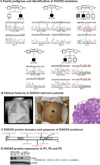
Figure 1
Figure 1. Identification of Mutations in the Dedicator of Cytokinesis 2 (DOCK2) Gene in Patients with Combined Immunodeficiency
Panel A shows the pedigrees of five families with affected individuals indicated by solid symbols and chromatograms corresponding to the identified DOCK2 mutations in five studied patients (P1–P5) and heterozygous carriers for each family. Panel B illustrates the clinical spectrum of DOCK2 deficiency (from left to right): episode of pneumonia in P2 requiring intubation; photograph of the skin rash, showing the vesicular lesions due to chickenpox in P3; neutrophil infiltrate in colonic lamina propria and crypt epithelium consistent with focal active colitis in P4 (hematoxylin eosin staining, magnification ×100). Panel C shows the distribution of the identified mutations relative to the DOCK2 protein structure depicting the SRC homology 3 (SH3) domain, the DOCK homology region (DHR)-1 domain and the DHR-2 domain. Panel D shows the immunoblot analysis of protein lysates obtained from EBV-transformed B-cell lines derived from P3 and two healthy donors (HD) and protein lysates obtained from T-cell lines derived from P1, P2, and a healthy donor (HD). For panels D, GAPDH served as protein loading control.
Figure 2
Figure 2. Defective RAC1 Activation, Lymphocyte Migration and Actin Polymerization in DOCK2-deficient Patients
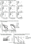
Panel A shows impaired RAC1 activation in T-cell lines from P1 and P2 as compared to a healthy donor (HD) and P2’s mother (P2M) upon stimulation of the T-cell receptor using anti-CD3 monoclonal antibodies, comparison done with one-way ANOVA. ***, p<0.001; **, p<0.01; horizontal bars represent means; error bars represent means ± s.d. of three independent experiments. Panel B shows reduced T- and B-cell migration in response to CCL21 or CXCL12 in P1 (grey bar) and P2 (black bar) as compared to a healthy control (white bar). NS, not stimulated. Panels C and D show reduced levels of polymerized, filamentous actin (F-actin) in P1 and P2 as revealed by phalloidin staining in T and B cells, respectively. MFI – mean fluorescence intensity.
Figure 3
Figure 3. NK Cell Cytotoxicity and Signaling Defects in DOCK2-deficient Patients
Panel A shows impaired NK-cell degranulation (monitored by CD107a surface expression) upon stimulation with K562 cells in P1 and P2 as compared to healthy donors (HD, n=10), horizontal bars represent means; error bars represent means ± s.d. Panel B shows defective degranulation upon triggering activating NK-cell receptors in P1 and P2 as compared to six healthy donors, horizontal bars represent means; error bars represent means ± s.d. n.d., not done. Panel C shows impaired actin polymerization upon triggering activating NK-cell receptors CD16 (left) and NKp46 (right). Panel D shows impaired MEK/ERK phosphorylation in P2’s NK cells upon triggering activating receptors, as detected by flow cytometry. Panel E shows decreased IFN-γ production upon P2’s NK cell stimulation with IL-12 and IL-18.
Figure 4
Figure 4. Impaired IFN Responses to Viruses in DOCK2-deficient Leukocytes and Fibroblasts
Panel A shows impaired IFN-α and IFN-β production by DOCK2-deficient peripheral blood mononuclear cells (PBMC) upon exposure to either herpes simplex virus 1 (HSV-1) or vesicular stomatitis virus (VSV) for 24 hours each. By contrast, production of IL-6 by the patients’ PBMCs was comparable to the healthy donor (HD) PBMCs, and served as assay control. NI, not infected. Panel B shows increased EMCV-induced cell death of DOCK2-deficient SV40-fibroblasts (left panel) and rescue by the addition of IFN-α2b (right panel). TLR3- and STAT1-deficient SV40-immortalized fibroblast cell lines served as examples of defective IFN-dependent anti-viral immunity. Panel C shows immunoblot analysis of protein lysates obtained from SV40-fibroblasts derived from a healthy donor (HD) and P2, as well as from P2’s mock-transduced and DOCK2-transduced fibroblasts. Panel D shows rescue of EMCV-induced cell death of P2 SV40-fibroblasts by exogenous expression of wild-type DOCK2. STAT1-deficient fibroblasts were used as negative control. The data in panels B and D depict one representative out of three independent experiments with technical triplcates carried out in each experiment. MOI, multiplicity of infection.

References

    1. Al-Herz W, Bousfiha A, Casanova JL, et al. Primary immunodeficiency diseases: an update on the classification from the international union of immunological societies expert committee for primary immunodeficiency. Front Immunol. 2014;5:162. - PMC - PubMed
    1. Notarangelo LD. Functional T cell immunodeficiencies (with T cells present) Annual review of immunology. 2013;31:195–225. - PubMed
    1. Koboldt DC, Steinberg KM, Larson DE, Wilson RK, Mardis ER. The next-generation sequencing revolution and its impact on genomics. Cell. 2013;155:27–38. - PMC - PubMed
    1. Conley ME, Casanova JL. Discovery of single-gene inborn errors of immunity by next generation sequencing. Curr Opin Immunol. 2014;30C:17–23. - PMC - PubMed
    1. Ng SB, Buckingham KJ, Lee C, et al. Exome sequencing identifies the cause of a mendelian disorder. Nat Genet. 2010;42:30–35. - PMC - PubMed

Publication types

MeSH terms

Substances