Clinical Trial
. 2015 Jun 17;10(6):e0123683.
doi: 10.1371/journal.pone.0123683. eCollection 2015.

Efficacy of Lapatinib in Therapy-Resistant HER2-Positive Circulating Tumor Cells in Metastatic Breast Cancer

Affiliations
Clinical Trial

Efficacy of Lapatinib in Therapy-Resistant HER2-Positive Circulating Tumor Cells in Metastatic Breast Cancer

Sofia Agelaki et al. PLoS One. .

Abstract

Background: To evaluate the efficacy of lapatinib, a dual EGFR and HER2 tyrosine kinase inhibitor, in therapy-resistant HER2-positive CTCs in metastatic breast cancer (MBC).

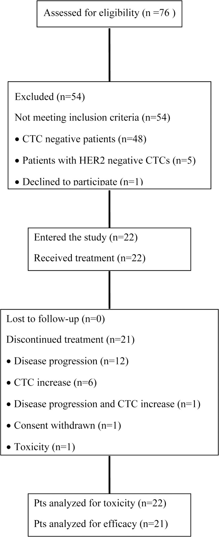
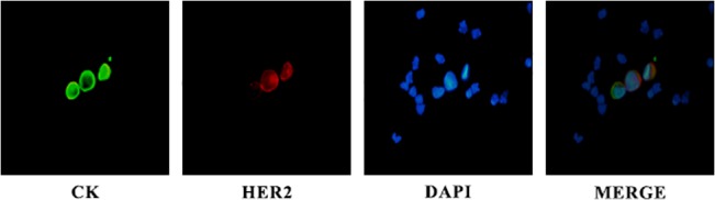
Patients and methods: Patients with MBC and HER2-positive CTCs despite disease stabilization or response to prior therapy, received lapatinib 1500 mg daily in monthly cycles, till disease progression or CTC increase. CTC monitoring was performed by immunofluorescent microscopy using cytospins of peripheral blood mononuclear cells (PBMCs) double stained for HER2 or EGFR and cytokeratin.

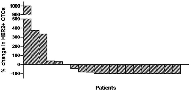
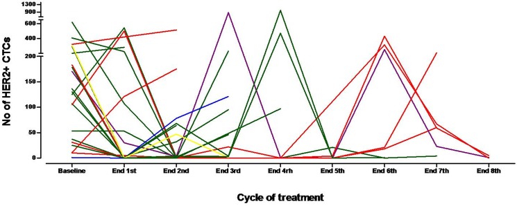
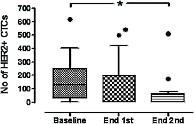
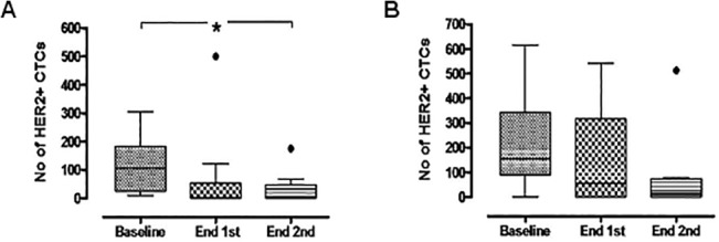
Results: A total of 120 cycles were administered in 22 patients; median age was 62.5 years, 15 (68.2%) patients were post-menopausal and 20 (90.1%) had HER2-negative primary tumors. At the end of the second course, HER2-positive CTC counts decreased in 76.2% of patients; the median number of HER2-positive CTCs/patient also declined significantly (p = 0.013), however the decrease was significant only among patients presenting disease stabilization (p = 0.018) but not among those with disease progression during lapatinib treatment. No objective responses were observed. All CTC-positive patients harbored EGFR-positive CTCs on progression compared to 62.5% at baseline (p = 0.054). The ratio of EGFR-positive CTCs/total CTCs detected in all patients increased from 17.1% at baseline to 37.6% on progression, whereas the mean percentage of HER2-negative CTCs/patient increased from 2.4% to 30.6% (p = 0.03).

Conclusions: The above results indicate that lapatinib is effective in decreasing HER2-positive CTCs in patients with MBC irrespectively of the HER2 status of the primary tumor and imply the feasibility of monitoring the molecular changes on CTCs during treatment with targeted agents.

Trial registration: Clinical trial.gov NCT00694252.


Conflict of interest statement

Competing Interests: This work was partly supported by a research grant from the Hellenic Society of Medical Oncology (HESMO), GlaxoSmithKline (GSK), and the Cretan Association for Biomedical Research (CABR). VG has received research grants and honorarium for participation in advisory boards from GSK, Amgen, Novartis and Sanofi. All other authors have declared that no competing interests exist. This does not alter the authors' adherence to PLOS ONE policies on sharing data and materials.

Figures

Fig 1
Fig 1. CONSORT flow diagram of the study.
Fig 2
Fig 2. A representative picture of CTCs detected on a patient’s cytospin.
Cells were double stained using A45 B/B3 (green), HER2 (red) antibodies, and DAPI (blue) and evaluated by immunofluorescence microscopy. Presented is a HER2-negative CTC in comparison with two HER2-positive CTCs.
Fig 3
Fig 3. Waterfall plot of the % change in HER2-positive CTC numbers in patients treated with lapatinib (n = 20) after the completion of the first treatment cycle as compared to the baseline; in one patient no information on CTC counts was available.
Fig 4
Fig 4. Patient-specific trajectories of HER2-positive CTCs during the first 8 cycles of treatment.
The trajectories of CTCs are marked red in patients that discontinued treatment due to increase in CTC counts (n = 6), green in cases with disease progression (n = 12) and blue for both CTC increase and disease progression (n = 1). One patient withdrew consent (depicted in yellow) and one was still on treatment at the time of the analysis (purple).
Fig 5
Fig 5. Box-plot of total HER2-positive CTC counts detected in the whole group of patients at baseline and after the completion of the first and second treatment cycles.
Shown are the median values ± SD of HER2-positive CTC numbers. HER2-positive CTCs declined from a median of 130 per patient (range 1–617), to a median of 2 (range 0–542) (p = 0.103) and 2 per patient (range 0–513) at the end of the first and second treatment courses, respectively (p = 0.013). Boxes represent the 25th and 75th percentile; line within the box shows the median value (Whiskers: Tukey).
Fig 6
Fig 6. Kaplan-Meier estimate of Progression-Free-Survival (PFS) in patients with metaststic breast cancer receiving lapatinib (n = 21).
Fig 7
Fig 7. Box-plot of total HER2-positive CTC counts detected in patients with stable disease (A) and progressive disease (B) as best response to treatment at baseline and after the completion of the first and second treatment cycles.
Shown are the median values ± SD of HER2-positive CTC numbers. In patients with stable disease CTCs declined from a median of 105 per patient (range 10–305) at baseline to 0 (range 0–500) and 0 (range 0–175), post-first (p = 0.193) and post-second cycles (p = 0.018), respectively. No significant differences in CTC counts were observed in patients with progressive disease. Boxes represent the 25th and 75th percentile; line within the box shows the median value (Whiskers: Tukey).

Similar articles

Cited by

References

    1. Husemann Y, Geigl JB, Schubert F, Musiani P, Meyer M, et al. (2008) Systemic spread is an early step in breast cancer. Cancer Cell 13: 58–68. 10.1016/j.ccr.2007.12.003 - DOI - PubMed
    1. Ignatiadis M, Georgoulias V, Mavroudis D (2008) Circulating tumor cells in breast cancer. Curr Opin Obstet Gynecol 20: 55–60. 10.1097/GCO.0b013e3282f22b2e - DOI - PubMed
    1. Braun S, Vogl FD, Naume B, Janni W, Osborne MP, et al. (2005) A pooled analysis of bone marrow micrometastasis in breast cancer. N Engl J Med 353: 793–802. - PubMed
    1. Stathopoulou A, Vlachonikolis I, Mavroudis D, Perraki M, Kouroussis C, et al. (2002) Molecular detection of cytokeratin-19-positive cells in the peripheral blood of patients with operable breast cancer: evaluation of their prognostic significance. J Clin Oncol 20: 3404–3412. - PubMed
    1. Xenidis N, Perraki M, Kafousi M, Apostolaki S, Bolonaki I, et al. (2006) Predictive and prognostic value of peripheral blood cytokeratin-19 mRNA-positive cells detected by real-time polymerase chain reaction in node-negative breast cancer patients. J Clin Oncol 24: 3756–3762. - PubMed

Publication types

MeSH terms

Associated data