. 2015 Sep;88(1053):20150099.
doi: 10.1259/bjr.20150099. Epub 2015 Jun 17.

Semi-quantitative contrast-enhanced MR analysis of indeterminate ovarian tumours: when to say malignancy?

Affiliations

Semi-quantitative contrast-enhanced MR analysis of indeterminate ovarian tumours: when to say malignancy?

S M Mansour et al. Br J Radiol. 2015 Sep.

Abstract

Objective: To evaluate the ability of dynamic post-contrast sequence to specify indeterminate ovarian masses with inconclusive MR features of malignancy. Since management is dramatically different, special focus on the ability to differentiate borderline from invasive malignancy was considered.

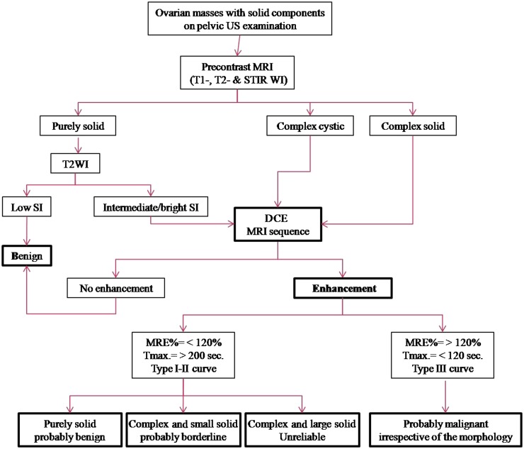
Methods: 150 ovarian masses were detected by pelvic ultrasound in 124 patients. Masses had been considered for dynamic post-contrast MRI. We expressed the kinetic parameters (i.e. enhancement amplitude, time peak of maximal uptake and maximal slope) in the form of maximum relative enhancement percentage (MRE%), time of maximal peak of contrast uptake (Tmax) and slope enhancement ratio (SER) curves. Histological findings were the gold standard of reference.

Results: Malignant ovarian masses showed higher MRE% than benign and borderline masses (p < 0.001). Tmax was shorter for malignant than benign (p < 0.01) and borderline (p < 0.001) ovarian masses. SER curves were the most suggestive of malignancy with a specificity and accuracy of 85.7% and 84.7%, respectively.

Conclusion: Dynamic contrast-enhanced MRI could be a specific sequence to differentiate ovarian masses with indeterminate MR morphology with a special discrimination for low potential from invasive ovarian malignancy.

Advances in knowledge: The study evaluated the diagnostic performance of the individual parameters of dynamic post-contrast MR sequence in evaluating ovarian masses. Management divert between benign, borderline and invasive malignant masses; our work presented a cut-off value for the peak of contrast uptake of 120%, which helped in the differentiation between benign and malignant tumours; the SER curves with Type III (early washout) pattern that was indicative of invasive malignancy was more specific than borderline malignancy.


Figures

Figure 1.
Figure 1.
An 18-year-old female with bilateral ovarian masses. Right luteinized thecoma and left simple cyst. (a) Coronal T2 weighted fast spin echo shows the uterus sandwiched between large anteriorly located pelvic solid mass of heterogeneous high signal intensity (SI) and simple cyst related to the left ovary seen at the cul-de-sac. Note the presence of ascetic fluid at the right iliac region. (b) Presence of high SI areas in the periphery of the mass on axial T1 weighted spectral pre-saturation inversion recovery image suggestive of haemorrhagic infarction. (c) Early and delayed post-contrast T1 high-resolution isotropic volumetric examination images show delayed contrast uptake of the mass with no appreciable enhancement at the infracted periphery. Kinetics was initial enhancement peak at 154 s with corresponding maximum relative enhancement percentage of 154% and Type I benign curve pattern. A, ascites; L, left; R, right; U, uterus.
Figure 2.
Figure 2.
A 55-year-old female presented with right ovarian borderline serous cystadenoma. (a) Sagittal T2 weighted fast spin echo shows large complex cystic, mass (M; curved arrows) with septations and posterior wall-based cauliflower soft tissue (straight arrow). Note the vaginal prolapse and intussusception of the cervix. (b) A collective figure: the left column represents sagittal post-contrast T1 high-resolution isotropic volumetric examination image and the colour mapping images (that could detect the most vascular portion of the tumour). The right column represents the kinetic analysis of delayed initial peak of contrast uptake at 288 s with corresponding maximum relative enhancement percentage of 87% and Type I (benign) curve pattern. The morphological features were in favour of invasive malignancy, yet the post-contrast dynamic parameters were more towards benign kinetics. The latter finding was explained by the tumour pathology being a borderline tumour. R, rectum; U, uterus; UB, urinary bladder.
Figure 3.
Figure 3.
A 54-year-old female patient with right ovarian borderline cystadenofibroma. (a) Sagittal T2 weighted fast spin echo shows complex ovarian mass with small solid component (blue arrows). Note the marked cervistis in the form of multiple nabothian cysts. (b) Three-dimensional sagittal oblique multiplanar reformatting reconstructed post-contrast image shows the right ovarian mass and the uterus along its whole length, the solid component of the mass displayed uptake in a comparable timing to the uterine myometrium. (c) Semi-quantitative parameters display delayed Tmax at 238 s, maximum relative enhancement percentage of 87% and Type I progressively rising curve pattern. The suspicious complex features of the ovarian mass and the age of the patient favour invasive malignant pathology, yet the kinetics were towards benign neoangiogenesis that coincided with the pathology being a borderline mass. C, cervix; U, uterus; UB, urinary bladder. For colour image see online.
Figure 4.
Figure 4.
A post-menopausal nulliparous 45-year-old female with right ovarian squamous cell carcinoma arising on the top of immature cystic teratoma. (a) Sagittal T2 weighted fast spin echo shows large adnexal complex mass with rounded matted tuft of hair seen centred on fluid sedimentation levelling (black star). Associate mural-based lobulated soft-tissue component (white arrow). (b) A collective figure included in the upper row from left to right: sagittal post-contrast T1 high-resolution isotropic volumetric examination (source) image, subtraction post-contrast image (best distinction of the enhancing soft tissue seen adherent to the posterior wall) and colour-coded image. The lower row represented the kinetic analysis of early initial peak of contrast uptake at 78 s with corresponding maximum relative enhancement percentage of 112% and Type III malignant curve pattern. The last image in the lower row represented a colour mapping image (rapid and strongly enhancing areas are displayed in red or yellow, while areas of slow or weak enhancement appear green). U, uterus; UB, urinary bladder. For colour image see online.
Figure 5.
Figure 5.
Right ovarian poorly differentiated Sertoli–Leydig tumour in a 44-year-old female, amenorrhoeic since 7 months. (a) Coronal T2 weighted fast spin echo shows right ovary purely solid pelvic mass of intermediate signal intensity (star). Note that the right ovary shows few follicles. Normal left ovary (black arrow). (b) Quantitative assessment of the right ovarian mass displayed a high maximum relative enhancement percentage of 418%, Tmax of 119 s, and Type III malignant curve pattern seen demonstrated in a collective figure that included T1 high-resolution isotropic volumetric examination source image, subtraction, colour-coded and colour mapping images. C, cervix; R, rectum.
Figure 6.
Figure 6.
A 44-year-old female patient with right ovarian granulosa cell tumour. (a) Sagittal (right) and coronal (left) T2 weighted fast spin echo shows large pelvic predominantly solid mass (M) of intermediate to high signal intensity. Some scattered tiny cysts are seen within. (b) Three-dimensional axial oblique MPR reconstructed post-contrast image shows the large right ovarian mass, the uterus (U), right iliac region ascites (A), the normal left ovary and the rectum (R), all in one image. (c) Kinetic analysis of the right ovarian mass displays early Tmax at 78 s with corresponding maximum relative enhancement percentage of 213% and Type III malignant curve pattern. Asc., ascites; K, kidney; Lt., left; Rt., right; U, uterus; UB, urinary bladder.
Figure 7.
Figure 7.
A suggested MRI algorithm in the assessment of indeterminate ovarian masses with solid components detected on pelvic US examination. DCE, dynamic contrast-enhanced; MRE%, maximum relative enhancement percentage; SI, signal intensity.

References

    1. Jung SE, Lee JM, Rha SE, Byun JY, Jung JI, Hahn ST. CT and MR imaging of ovarian tumours with emphasis on differential diagnosis. Radiographics 2002; 22: 1305–25. doi: 10.1148/rg.226025033 - DOI - PubMed
    1. Berek JS, Hacker NF. Practical gynecologic oncology. 4th edn. Philadelphia, PA: Lippincott Williams & Wilkins; 2005. pp. 443–54.
    1. Berek JS, Hacker NF. Staging and second-look operations in ovarian cancer. In: Alberts DS, Surwit EA, eds. Ovarian cancer. Boston, MA: Martinus Nijhoff; 1985. pp. 109–27.
    1. Gultekin E, Gultekin OE, Cingillioglu B, Sayhan S, Sancil M, Yildirim Y. The value of frozen section evaluation in the management of borderline ovarian tumours. J Cancer Res Ther 2011; 7: 416–20. doi: 10.4103/0973-1482.92005 - DOI - PubMed
    1. Gupta N, Rajpal T, Sharma S. Evaluating the accuracy of frozen section in borderline ovarian tumours. J Clin Oncol 2013; 31(Suppl.): Abstract 5564.