. 2015 Jun 17;10(6):e0128443.
doi: 10.1371/journal.pone.0128443. eCollection 2015.

Control of Protein Activity and Cell Fate Specification via Light-Mediated Nuclear Translocation

Affiliations

Control of Protein Activity and Cell Fate Specification via Light-Mediated Nuclear Translocation

Hayretin Yumerefendi et al. PLoS One. .

Abstract

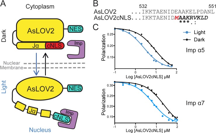
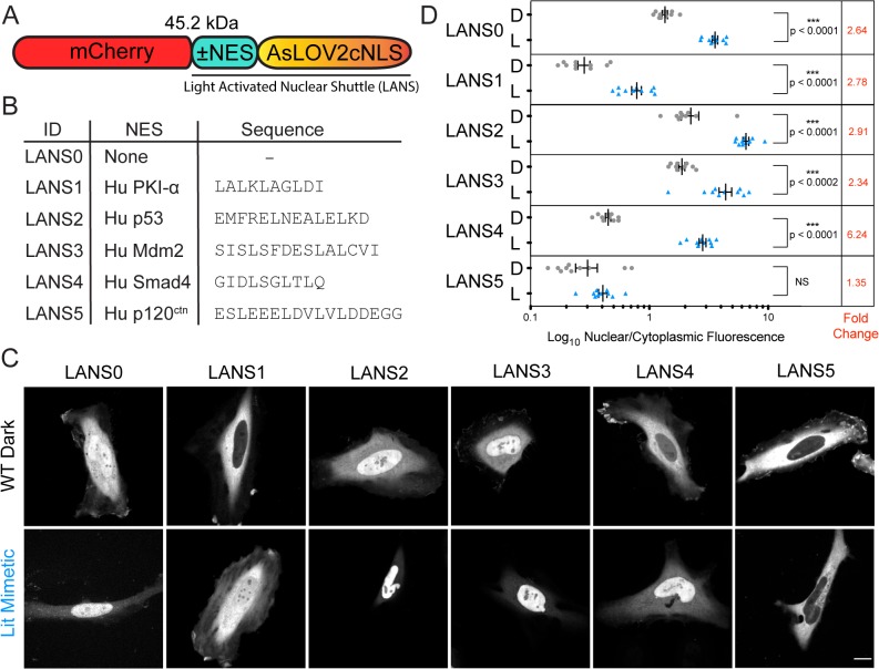
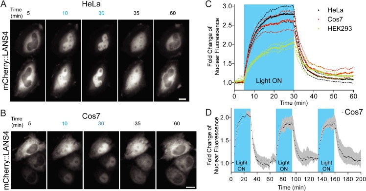
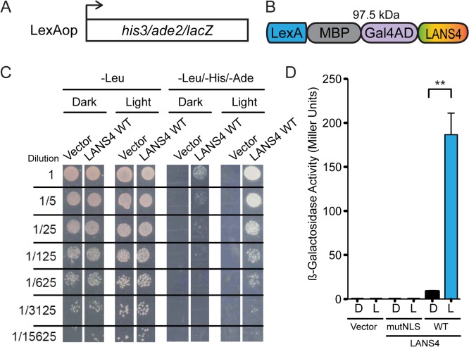
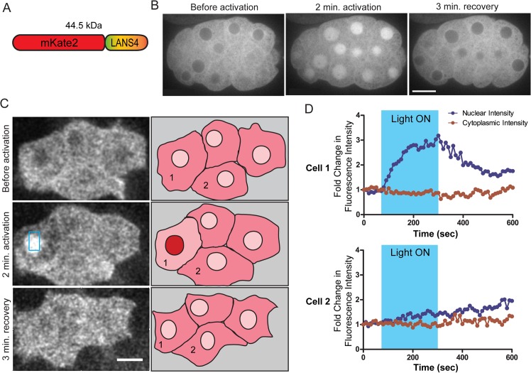
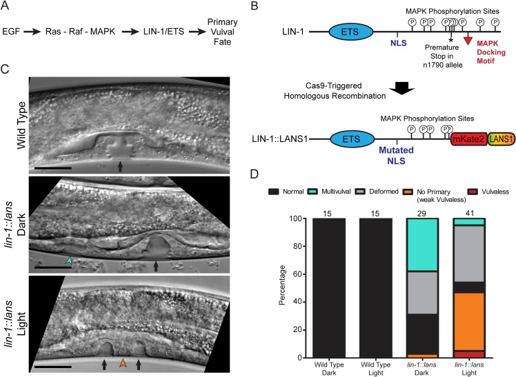
Light-activatable proteins allow precise spatial and temporal control of biological processes in living cells and animals. Several approaches have been developed for controlling protein localization with light, including the conditional inhibition of a nuclear localization signal (NLS) with the Light Oxygen Voltage (AsLOV2) domain of phototropin 1 from Avena sativa. In the dark, the switch adopts a closed conformation that sterically blocks the NLS motif. Upon activation with blue light the C-terminus of the protein unfolds, freeing the NLS to direct the protein to the nucleus. A previous study showed that this approach can be used to control the localization and activity of proteins in mammalian tissue culture cells. Here, we extend this result by characterizing the binding properties of a LOV/NLS switch and demonstrating that it can be used to control gene transcription in yeast. Additionally, we show that the switch, referred to as LANS (light-activated nuclear shuttle), functions in the C. elegans embryo and allows for control of nuclear localization in individual cells. By inserting LANS into the C. elegans lin-1 locus using Cas9-triggered homologous recombination, we demonstrated control of cell fate via light-dependent manipulation of a native transcription factor. We conclude that LANS can be a valuable experimental method for spatial and temporal control of nuclear localization in vivo.


Conflict of interest statement

Competing Interests: The authors have declared that no competing interests exist.

Figures

Fig 1
Fig 1. Design and biophysical characterization of light conditioned nuclear localization signal.
(A) Schematic of the Light Activated Nuclear Shuttle (LANS) design for light activated nuclear import (B) Sequence alignment of the wild type AsLOV2 and the designed AsLOV2cNLS (sequence identity and homology is marked according to CLUSTALW scheme). (C) Fluorescence polarization competitive binding assay of AsLOV2cNLS against human importin α5 and importin α7.
Fig 2
Fig 2. Confocal microscopy of LANS in HeLa cells.
(A) Schematic of the LANS constructs (B) List of the nuclear export signals tested. (C) Representative nuclear optical slices of cells used for the quantification of the nuclear/cytoplasmic distribution of the switch (scale bar = 15 μm). (D) Quantification of the effect of the nuclear export signal on the nuclear/cytoplasmic distribution of LANS (D—wild type construct imaged in the dark, L—lit mimetic I539E). Mean is reported ±SEM and statistical significance calculated with unpaired two-tailed t-students test; NS—Not Significant.
Fig 3
Fig 3. Real time light induced nuclear translocation of LANS4 in mammalian tissue culture cells.
(A) Representative images for light activation and reversion in HeLa cells and Cos7 (B) (scale bar = 25 μm); (c) Plotting the fold change of nuclear accumulations in HeLa, Cos7 and HEK293 (n = 4 each, mean reported ± SEM with dashed line). See also S1, S2 and S3 Movies. The blue shaded region indicates pulsed blue light activation (see Supplemental experimental procedures). (C) Multple activation reversion cycles in Cos7 (n = 2, mean reported ± SEM with shaded grey area). The blue shaded regions indicate pulsed blue light activation.
Fig 4
Fig 4. Light induced transcription via light mediated nuclear translocation in yeast.
(A) NMY51 contains his3, ade2 and lacZ genomic reporter genes under the control of LexAop. (B) Schematic of the LANS controlled artificial transcription factor in yeast (C) Growth assay of LANS controlled transcription factor in NMY51. The left panel shows growth on media lacking leucine, which confers plasmid resistance and demonstrates that the light used does not affect regular yeast growth. The right panel demonstrates light dependent growth on media lacking leucine, histidine and adenine. (D) β-galactosidase activity measurements upon blue light induced transcription activation with LANS4 n = 3 each, mean reported ± SEM and statistical significance is calculated with unpaired two-tailed t-student’s test (p = 0.0019).
Fig 5
Fig 5. Light activated nuclear translocation in C. elegans embryo.
(A) Schematic of the mKate2::LANS construct that was expressed in C. elegans embryos (B) Confocal images of an embryo expressing mKate2::LANS ubiquitously and subjected to photoactivation with blue light. Scale bars represent 10 μm. See also S4 Movie. (C) Left: Confocal images of four mKate2::LANS expressing MS lineage cells on the ventral surface of a late gastrulation-stage embryo. The blue box in the center image indicates the region that was photoactivated with blue light. Brightness and contrast were adjusted to compensate for photobleaching. Scale bar represents 5 μm. Right: Sketches summarizing the observed localization. Numbers correspond to the cell numbers in (D). See also S5 Movie. (D) Quantification of nuclear and cytoplasmic fluorescence intensities as a function of time for the two cells labelled in (C). Cell 1 was illuminated with blue light, and Cell 2 is a neighboring cell. These measurements were corrected for photobleaching (see materials and methods).
Fig 6
Fig 6. Control of vulval development via photoactivatable LIN-1.
(A) Simplified schematic of the role of LIN-1 in vulval fate specification. (B) Top: Schematic of the wild type LIN-1 protein. Bottom: Schematic of the LIN-1::LANS protein produced after modification of the native lin-1 locus using Cas9-triggered homologous recombination. See also S4 Fig. (C) DIC Images of the developing vulvae in mid-L4 larvae from the indicted strains and conditions. Top panel: Black arrow indicates the normal, symmetric vulval invagination. Middle panel: Black arrow indicates the main vulval invagination, and green arrowhead indicates an extra vulval invagination. Bottom panel: Orange arrowheads indicate small invaginations produced by the secondary vulval precursors, and black arrow indicates the plug of tissue derived from the failed primary cell. Scale bars represent 20 μm. (D) Quantification of phenotypes in the indicated strains and conditions. See Methods for detailed definitions of each phenotype. Numbers at the top of each bar indicate the total number of animals scored in this experiment. These data are from a single experiment; the experiment was repeated three times, using two independently isolated lin-1::lans alleles, with similar results.

References

    1. Cartwright P, Helin K. Nucleocytoplasmic shuttling of transcription factors. Cell Mol Life Sci. 2000;57(8–9):1193–206. - PMC - PubMed
    1. Eilers M, Picard D, Yamamoto KR, Bishop JM. Chimaeras of myc oncoprotein and steroid receptors cause hormone-dependent transformation of cells. Nature. 1989;340(6228):66–8. - PubMed
    1. Feil R, Brocard J, Mascrez B, LeMeur M, Metzger D, Chambon P. Ligand-activated site-specific recombination in mice. Proc Natl Acad Sci U S A. 1996;93(20):10887–90. - PMC - PubMed
    1. Metzger D, Clifford J, Chiba H, Chambon P. Conditional site-specific recombination in mammalian cells using a ligand-dependent chimeric Cre recombinase. Proc Natl Acad Sci U S A. 1995;92(15):6991–5. - PMC - PubMed
    1. Haruki H, Nishikawa J, Laemmli UK. The anchor-away technique: rapid, conditional establishment of yeast mutant phenotypes. Mol Cell. 2008;31(6):925–32. 10.1016/j.molcel.2008.07.020 - DOI - PubMed

Publication types

MeSH terms