. 2015 Jun 25;522(7557):497-501.
doi: 10.1038/nature14560. Epub 2015 Jun 17.

New cofactor supports α,β-unsaturated acid decarboxylation via 1,3-dipolar cycloaddition

Affiliations

New cofactor supports α,β-unsaturated acid decarboxylation via 1,3-dipolar cycloaddition

Karl A P Payne et al. Nature. .

Abstract

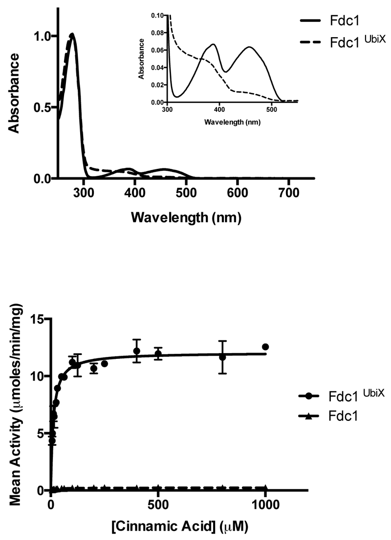
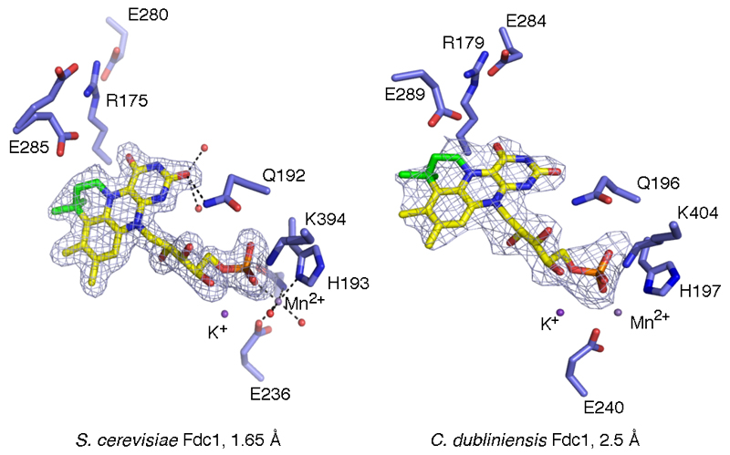
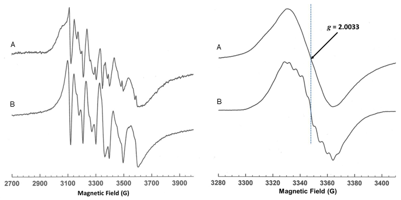
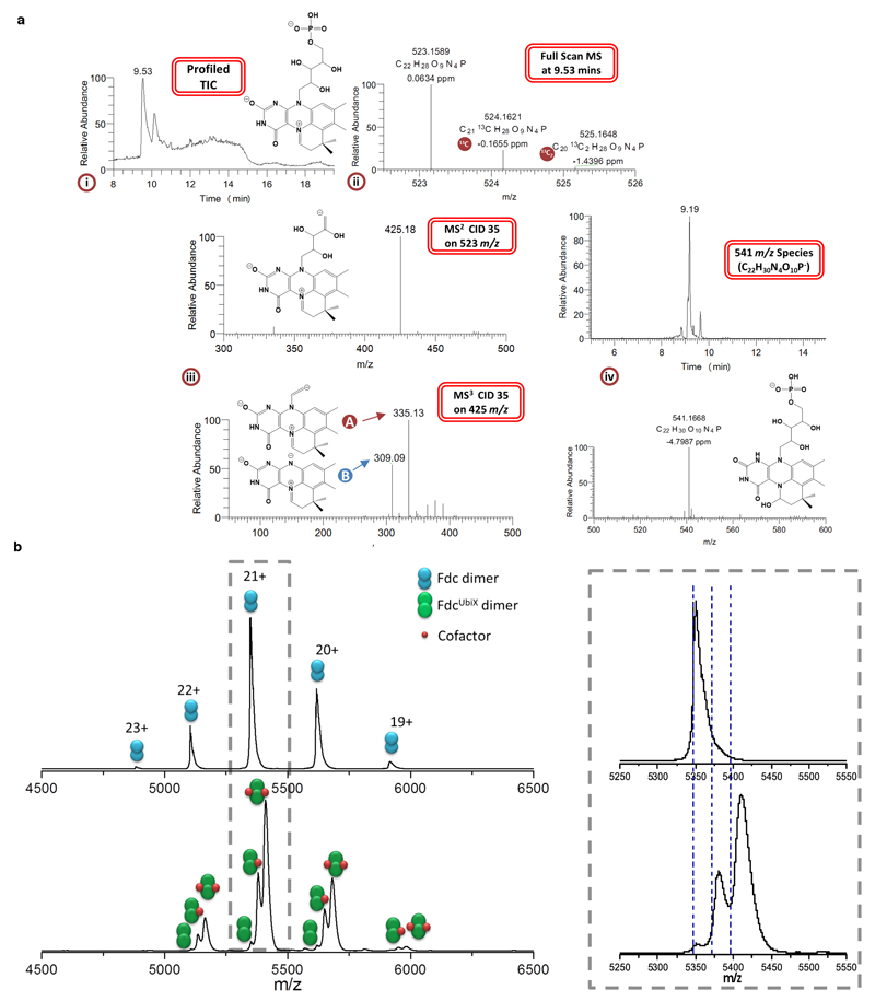
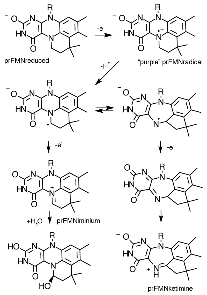
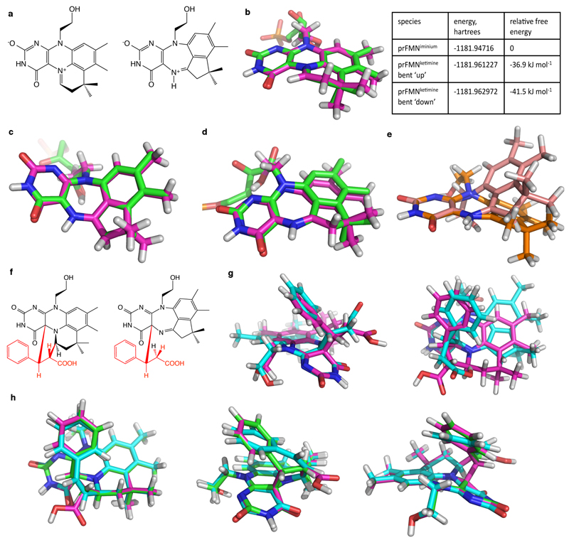
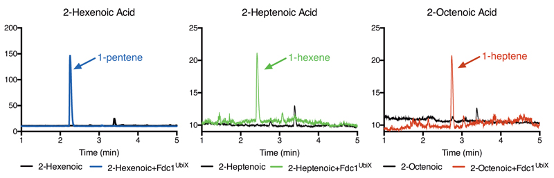
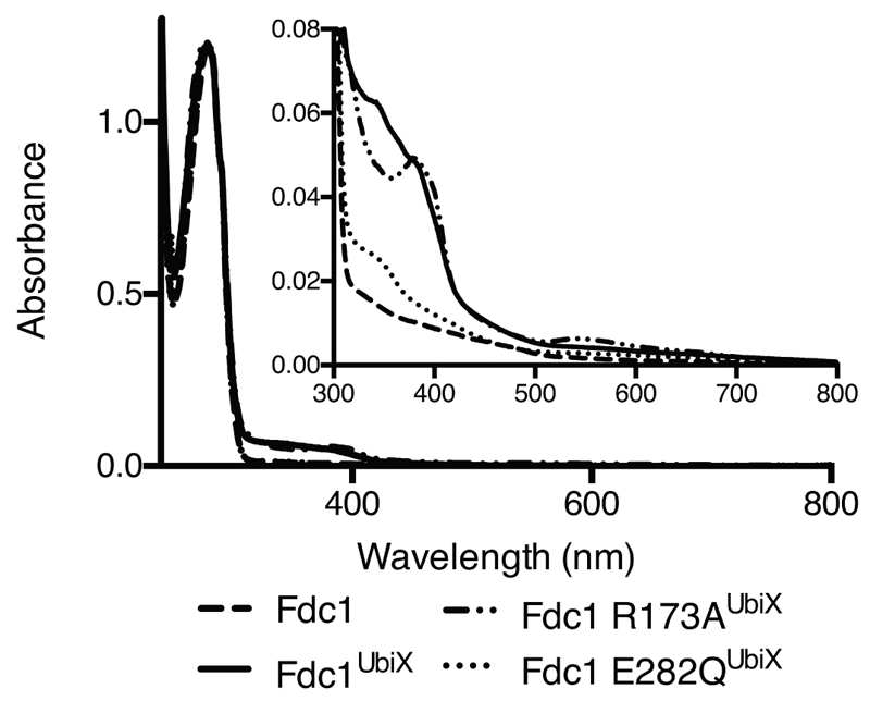
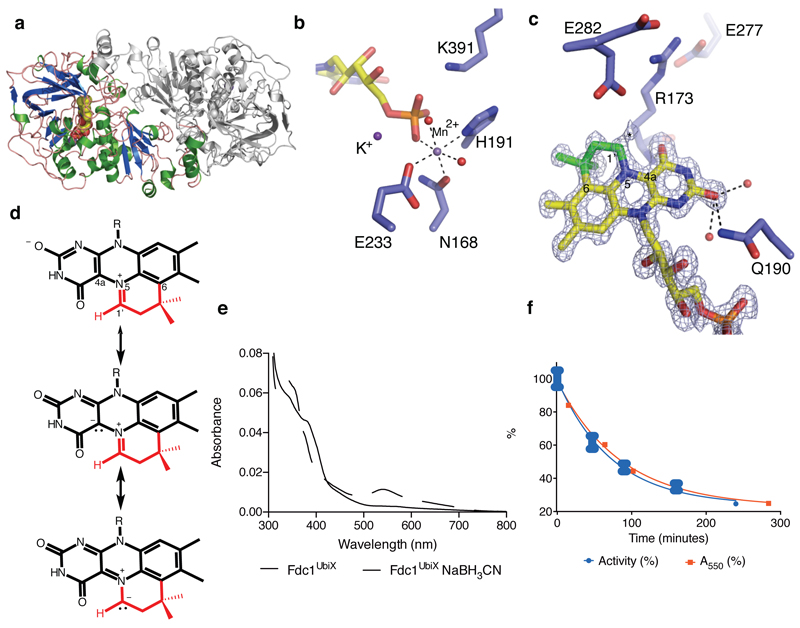
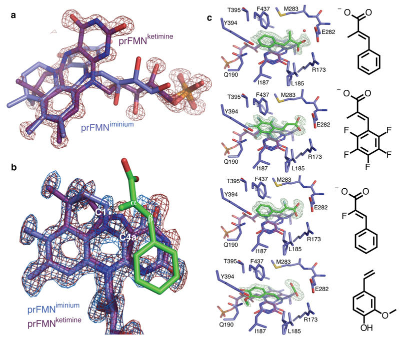
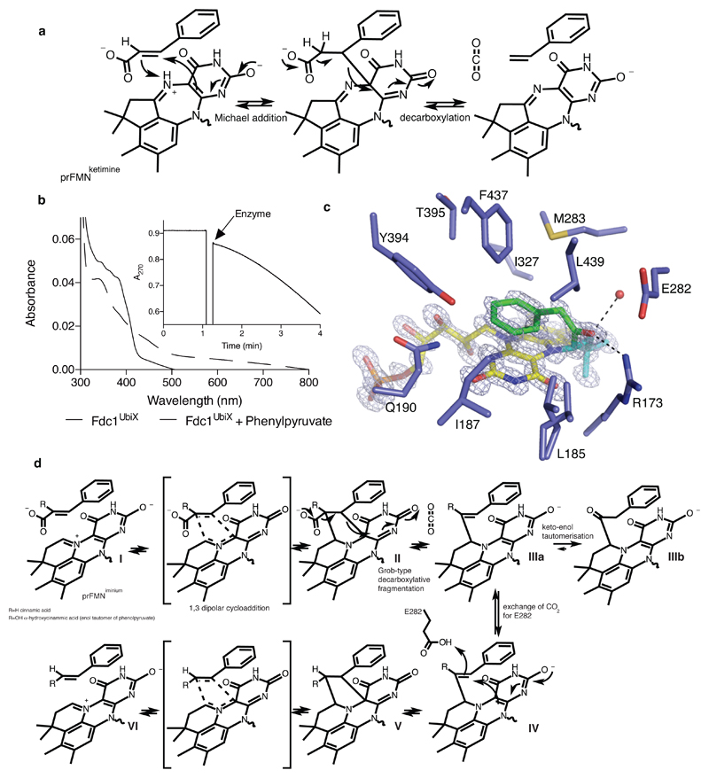
The bacterial ubiD and ubiX or the homologous fungal fdc1 and pad1 genes have been implicated in the non-oxidative reversible decarboxylation of aromatic substrates, and play a pivotal role in bacterial ubiquinone (also known as coenzyme Q) biosynthesis or microbial biodegradation of aromatic compounds, respectively. Despite biochemical studies on individual gene products, the composition and cofactor requirement of the enzyme responsible for in vivo decarboxylase activity remained unclear. Here we show that Fdc1 is solely responsible for the reversible decarboxylase activity, and that it requires a new type of cofactor: a prenylated flavin synthesized by the associated UbiX/Pad1. Atomic resolution crystal structures reveal that two distinct isomers of the oxidized cofactor can be observed, an isoalloxazine N5-iminium adduct and a N5 secondary ketimine species with markedly altered ring structure, both having azomethine ylide character. Substrate binding positions the dipolarophile enoic acid group directly above the azomethine ylide group. The structure of a covalent inhibitor-cofactor adduct suggests that 1,3-dipolar cycloaddition chemistry supports reversible decarboxylation in these enzymes. Although 1,3-dipolar cycloaddition is commonly used in organic chemistry, we propose that this presents the first example, to our knowledge, of an enzymatic 1,3-dipolar cycloaddition reaction. Our model for Fdc1/UbiD catalysis offers new routes in alkene hydrocarbon production or aryl (de)carboxylation.


Conflict of interest statement

The authors declare no competing financial interest.

Figures

Extended Data Fig 1
Extended Data Fig 1
HPLC chromatogram demonstrating enzymatic carboxylation of styrene by A. niger Fdc1UbiX. Chromatogram of a 10 mM styrene and saturating NaHCO3 solution incubated in the absence (blue) and presence (red) of the A. niger Fdc1UbiX enzyme.
Extended Data Fig 2
Extended Data Fig 2
S. cerevisiae FdcUbiX solution data. The left panel is a direct comparison to main Fig 1b. In this case, the non-UbiX coexpressed Fdc1 binds FMN weakly. The right panel shows S. cerevisiae FdcUbiX steady state kinetic data obtained with cinnamic acid. Error bars are s.e.m. n=3
Extended Data Fig 3
Extended Data Fig 3
A detailed view of the S. cerevisiae and C. dubliniensis FdcUbiX active site. The omit map corresponding to the prFMN cofactor is shown in a blue mesh contoured at 3 sigma. Atoms derived from dimethylallyl phosphate are shown in green. Both FdcUbiX crystals appeared colourless as observed for A. niger Fdc1UbiX, indicating neglible FMN binding.
Extended data Fig 4
Extended data Fig 4
Left panel. X-band continuous wave frozen solution EPR spectra of a) Fdc1 and b) Mn(H2O)62+ showing the characteristic six line pattern arising from the ms = ± ½ spin manifold of the S = 5/2 Mn2+ ion. The six line pattern reflects hyperfine coupling to the I = 5/2 55Mn nucleus and is sensitive to the environment of the ion, thus the differences between a) and b) indicate binding of Mn2+ to the enzyme.. Experimental conditions: microwave power 0.5 mW, modulation amplitude 7 G, temperature 20 K. Right panel. X-band continuous wave frozen solution EPR spectra of a) FdcUbiX reduced using sodium cyanoborohydride (NaBH3CN) and subsequently exposed to air. b) WT UbiX + DMAP reduced with dithionite and reoxidised with oxygen. Experimental conditions: microwave power 10 μW, modulation amplitude 1.5 G, temperature 20 K. Re-reduction using NaBH3CN of the modified cofactor formed in FdcUbiX (prFMNiminium)gives rise to a radical on air oxidation with the same g value (gav) and line width as that formed on the modified flavin (prFMN) in UbiX. However, the lack of the distinctive fine structure in a) that is normally observed for the UbiX radical, b), suggests heterogeneity in the FdcUbiX radical or possibly a magnetic interaction with the nearby Mn2+ ion in Fdc.
Extended Data Figure 5
a (i) Structural elucidation of the reduced UbiD/Fdc1 co-factor. Full scan TIC created under a gradient elution using H2O/ acetonitrile both containing 0.1% formic acid indicating a major peak apex at 9.53 mins with a 52/48 solvent composition. Also shown is the proposed structure of 523 m/z. Mass spectrum taken at major peak apex in (ii) (9.53 mins) indicating an associated full scan molecular ion peak with m/z = 523.1589 (M+ = C22H28N4O9P) at a resolution of 58501 with a mass accuracy of 0.06 ppm. Also eluting alongside the target molecule are two isotopic variants containing 13C and 13C2. The 13C peak is displaying a relative abundance of 22 to the 523m/z peak which is in line with the number of carbon atoms contained within the structure. Fragmentation of the 523.16 m/z molecular ion peak (iii) in an automated data dependent manner using helium based-chemical induced dissociation (CID level 35) generated a spectral tree that indicates the removal of the phosphate head group at the MS2 level generating 425.18 m/z. Subsequent MS3 level activation (CID 35) on 425.18 m/z partially (A) or completely (B) removing the tail group from the newly formed 4-ring system generating 335.13 m/z or 309.09 m/z, respectively. The presence of an oxidised species with 541 m/z is also reported (iv). b Native mass spectra of Fdc (top) and FdcUbiX (bottom). Fdc presents in charge states 19+ to 23+ while FdcUbiX in charge states 19+ to 22+. Right hand spectrum; an enlarged view of the 21+ charge state. The predicted masses are shown by blue dashed lines. The spectrum of FdcUbiX shows that approximately two thirds of the ions have 2 non-covalently bound cofactors, approximately one third have one non-covalently bound cofactor and there is a small amount with no cofactor bound. Fdc contains no cofactor. The measured mass of the Fdc dimer is 112 265 Da (predicted mass from sequence is 112 270 Da). The measured mass for the apo form of FdcUbiX is 112 345 Da, slightly higher than for Fdc which is attributed to an increased retention of salt. The mass difference of +80 Da corresponds to the mass of 2 potassium adducts. For the FdcUbiX species with one bound cofactor, the measured mass (112 968 Da) is 178 Da higher than predicted. The predicted mass corresponds to the left hand side of the peak which is the protein+cofactor with no extra salt retained. The extra mass could be attributed to 2 Mn2+ ions and 2 K+ ions. The FdcUbiX bound to 2 cofactors has a measured mass of 113 583 Da which is 268 Da larger than expected. Again, however, the predicted mass corresponds to the left hand side of the peak. These spectra indicate that the protein dimer carries either 1 or 2 cofactors of 525 Da, along with a variety of other salt adducts. The extent of adductation is higher for FdcUbiX and increases with bound co-factors, indicating that the addition of the cofactor recruits counter ions.
Extended Data Fig 6
Extended Data Fig 6
A proposed mechanism for cofactor maturation in FdcUbix through oxidation. We propose the prFMNreduced cofactor produced by UbiX is bound by apo-Fdc and oxidized in a stepwise manner. While the initial radical species resembles that observed during non-physiological oxidation of the prFMNreduced:UbiX product complex, we proposed that in the Fdc enzyme proton abstraction from the prFMN radical C1’ leads to a distinct radical species. The latter can either be oxidized further to the corresponding prFMNiminium or, via a radical based isomerisation process, form the central seven-membered ring ultimately leading to prFMNketimine.
Extended Data Fig 7
Extended Data Fig 7
DFT models of the proposed prFMNiminium and prFMNketimine isomers and their substrate adducts. a) Chemical structures of the prFMNiminium and prFMNketimine models. DFT models (pink carbons) are overlaid with crystal coordinates (green carbons) of prFMNiminium b) and the two butterfly bent conformations of prFMNketimine c) and d). The substrate-free (bent ‘up’) prFMNketimine is shown in c) and the more planar substrate-bound (bent ‘down’) in d). For comparison of the extent of the butterfly bending of prFMNketimine, the two DFT models in c) and d) are aligned over the 4 ring nitrogen atoms and overlaid in e). f) Chemical structures of the proposed initial prFMNiminium and prFMNketimine substrate adducts with the cinnamic acid substrate highlighted in red. The DFT-optimised structures of these species are overlaid in g) with the prFMNiminium species shown with pink carbons and the prFMNketimine species with teal carbons. Three projections of overlaid DFT models of the prFMNiminium species with substrate bound (pink carbons; reproduced from g), after substrate decarboxylation (teal carbons; substrate double bond is cis) and upon protonation of the substrate beta carbon (green carbons) are shown in h). Note that the substrate carboxylate was artificially protonated in these models to maintain charge neutrality. Models were geometry optimised in the gas phase using either the B3LYP/6-311++G(d,p) (panels a-e) or BH&H/6-311++G(d,p) (panels f-h)_level of theory. BH&H was chosen over B3LYP for the substrate adducts as BH&H has been shown to better describe pi-stacking interactions, which are likely to occur between the modified isoalloxazine and substrate phenyl moieties. Harmonic vibrational frequencies calculated using normal mode analysis were used to confirm that optimised geometries of all species were in local or global minima. Absolute energies and relative free energies of the substrate-free species, determined from the normal mode calculations, are given in the table, top right. Cartesian coordinates of the optimised structures are given in Supplementary Information.
Extended Data Fig 8
Extended Data Fig 8
Gas chromatogram showing products formed from a solution of respectively 10 mM 2-hexenoic (1-pentene, blue; enzyme free control in black ), 10 mM 2-heptenoic (1-hexene, green; enzyme free control in black ) or 10 mM 2-octeneoic acid (1-heptene, black; enzyme free control in green). No product (1-octene, red) could be detect from 10 mM 2-nonenoic acid. Identification and quantification of 1-alkenes was based on known standards.
Extended Data Fig 9
Extended Data Fig 9
UV-vis spectra of Fdc1 (614 mM), Fdc1UbiX (492 mM), Fdc1 R173AUbiX (749 mM) and Fdc1 E282QUbiX (171 mM) normalised on the A280 peak. Inset, close up of the additional spectral features present in the 300 – 500 nm region.
Fig 1
Fig 1. Fdc solution data.
a) Schematic overview of the reaction catalysed by Fdc/Pad and the related UbiD/UbiX proteins. b) UV-Vis spectra obtained for heterologous expressed A. niger Fdc, with and without co-expression of a ubiX gene. c) UV-vis observation of the enzymatic conversion of cinnamic acid to styrene via decarboxylation by FdcUbiX. The initial spectrum of cinnamic acid shows a λmax of 270 nm. Over time successive spectra show reduction of the 270 nm peak and appearance of a peak at 245 nm corresponding to styrene formation. d) Steady state kinetic parameters obtained for FdcUbiX for sorbic acid and a variety of cinnamic acid-type compounds (error bars are s.e.m. n=3).
Fig 2
Fig 2. FdcUbiX crystal structures.
a) Crystal structure of the A. niger FdcUbiX. The monomer present in the asymmetric unit is depicted in cartoon format (helices in green, sheets in blue) while the symmetry related monomer forming the FdcUbiX dimer is shown in grey. b) Detailed view of Mn2+ binding site, linking the modified FMN phosphate group to the protein. c) Detailed view of the modified FMN cofactor. Omit electron density map corresponding to the bound cofactor contoured at 5 sigma. A 4th ring (non-isoalloxazine derived atoms are shown in green) can clearly be observed. An asterisk indicates the position of additional weak electron density at the C1 position, that can be accounted for by partial hydrolysis. Key residues involved in polar interactions with the isoalloxazine derived cofactor moiety are shown in sticks. d) Chemical structure of the modified FMN bound by A. niger FdcUbiX as derived from atomic resolution density and high resolution mass spectrometry [Ext data Fig 5]. Atoms derived from dimethylallylmonophosphate are shown in red. The azomethine ylide resonance form is shown below. e) Oxidation of cyanoborohydride inactivated FdcUbiX leads to formation of a purple species, similar in UV-Vis and EPR spectral properties [Ext Data Fig 4] to that observed for the oxidized UbiX-prFMN complex. f) Following purification, FdcUbiX activity gradually decreases when incubated on ice, and formation of the purple prFMN radical following NaBH3CN treatment follows a similar trend (error bars are s.d. n=3).
Fig 3
Fig 3. FdcUbiX cofactor structure and ligand complexes.
a) Omit electron density map corresponding to a distinct isomer of the prFMN contoured 5 sigma. An expansion of the central ring of the isoalloxazine system can clearly be observed, with the distinct butterfly-bent conformation accompanied by altered conformation of the ribityl moiety. b) Detailed view of prFMNox isoforms in the FdcUbiX-substrate complexes. The omit electron density maps corresponding respectively to the prFMNiminium (in blue) and prFMNketimine (in red) species each contoured 5 sigma are shown for the alpha-methyl-cinnamic acid complex. c) A series of FdcUbiX substrate/product complexes. Selected active site residues of FdcUbiX are shown in atom colour sticks, with the omit electron density contoured at 5 sigma corresponding to respectively alpha-methyl-cinnamic acid, pentafluorocinnamic acid, alpha-fluorocinnamic acid and 4-vinyl-guaiacol.
Fig 4
Fig 4. FdcUbiX mechanism.
a) Proposed mechanism for FdcUbiX catalysis using the prFMN ketimine form. b) Incubation of FdcUbiX with phenylpyruvate leads to changes in the UV-Vis spectrum. The inset shows conversion of cinnamic acid to styrene (monitored at 270nm) after addition of phenylpyruvate treated FdcUbiX. c) Active site of FdcUbiX in complex with a phenylpyruvate derived adduct. The omit electron density map (contoured at 7 sigma, in blue) corresponding the prFMNiminium-phenylpyruvate adduct is shown. Atoms derived from phenylpyruvate are depicted in green. d) Proposed mechanism for FdcUbiX catalysis and phenylacetaldehyde adduct formation of the prFMNiminium form.

Comment in

References

    1. Aussel L, Pierrel F, Loiseau L, Lombard M, Fontecave M, Barras F. Biosynthesis and physiology of coenzyme Q in bacteria. Biochim Biophys Acta. 2014;1837:1004–1011. - PubMed
    1. Gulmezian M, Hyman KR, Marbois BN, Clarke CF, Javor GT. The role of UbiX in Escherichia coli coenzyme Q biosynthesis. Arch Biochem Biophys. 2007;467:144–153. - PMC - PubMed
    1. Leppik RA, Young IG, Gibson F. Membrane-associated reactions in ubiquinone biosynthesis in Escherichia coli. 3-octoprenyl-4-hydroxybenzoate carboxy-lyase. Biochim Biophys Acta. 1976;436:800–810. - PubMed
    1. Erb TJ. Carboxylases in natural and synthetic microbial pathways. Appl Environ Microbiol. 2011;77:8466–8477. - PMC - PubMed
    1. Boll M, Loeffler C, Morris BEL, Kung JW. Anaerobic degradation of homocyclic aromatic compounds via arylcarboxyl-coenzyme A esters: organisms, strategies and key enzymes. Environ Microbiol. 2014;16:612–627. - PubMed

Publication types

MeSH terms