Review
. 2015 Jun 16;42(6):1005-19.
doi: 10.1016/j.immuni.2015.06.006.

Interleukin-33 in Tissue Homeostasis, Injury, and Inflammation

Affiliations
Review

Interleukin-33 in Tissue Homeostasis, Injury, and Inflammation

Ari B Molofsky et al. Immunity. .

Abstract

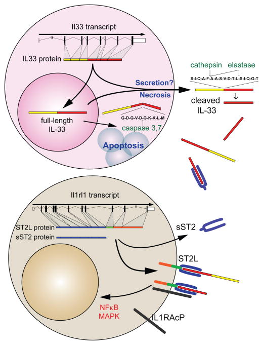
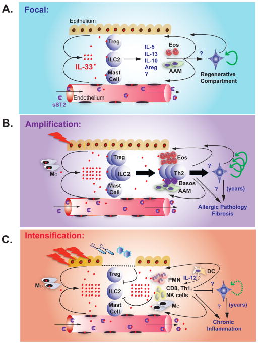
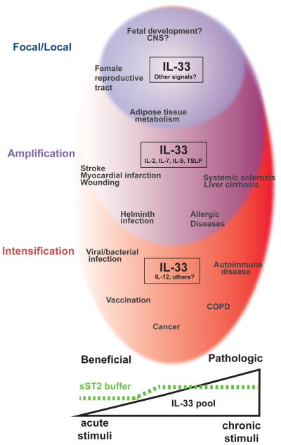
Interleukin-33 (IL-33) is a nuclear-associated cytokine of the IL-1 family originally described as a potent inducer of allergic type 2 immunity. IL-33 signals via the receptor ST2, which is highly expressed on group 2 innate lymphoid cells (ILC2s) and T helper 2 (Th2) cells, thus underpinning its association with helminth infection and allergic pathology. Recent studies have revealed ST2 expression on subsets of regulatory T cells, and for a role for IL-33 in tissue homeostasis and repair that suggests previously unrecognized interactions within these cellular networks. IL-33 can participate in pathologic fibrotic reactions, or, in the setting of microbial invasion, can cooperate with inflammatory cytokines to promote responses by cytotoxic NK cells, Th1 cells, and CD8(+) T cells. Here, we highlight the regulation and function of IL-33 and ST2 and review their roles in homeostasis, damage, and inflammation, suggesting a conceptual framework for future studies.


Conflict of interest statement

The authors have no conflicting financial interests.

Figures

Figure 1
Figure 1. IL-33-ST2 molecular characteristics
IL-33 is transcribed from seven coding exons and transported to the nucleus. During lytic cell death associated with necrosis or necroptosis, or possibly via direct secretion from intact cells, full-length IL-33 is released from the nucleus into the extracellular environment. Activation of apoptotic pathways leads to inactivation of IL-33 via caspase 3- or 7-mediated cleavage. Once released, full-length IL-33 can be further processed by serine proteases, such as cathepsin-G and elastase, into forms with increased activity. The IL-33 receptor ST2 is produced in two forms, a short soluble (sST2) or longer membrane-bound form (ST2L). sST2 is constitutively expressed, but can be induced during tissue damage, and binds IL-33 and restricts its availability. In contrast, both full-length and actively processed IL-33 bind to ST2L in combination with IL-1RAcP on target cells and induces canonical NFκB and MAPK signaling pathways leading to cellular activation and proliferation.
Figure 2
Figure 2. Progressive stages of IL-33-ST2
A. Homeostasis. IL-33 is present in the nuclei of a subset of epithelial and endothelial cells at rest. Poorly understood signals promote IL-33 release or focal cellular necrosis. IL-33 acts on tissue-resident ST2-expressing ILC2, Treg, and possibly mast cells, inducing the production of IL-5, IL-13, IL-10, amphiregulin (Areg), and other signals that promote eosinophils (Eos) and alternatively activated macrophages (AAM). These cells and signals feed back on the tissue and may regulate remodeling and limit inflammation, in part by activating tissue progenitor cells. sST2 in the blood prevents systemic IL-33 effects. B. Amplification. During tissue allergic insults and injury, epithelial and endothelial cells release IL-33, likely via necrosis, and IL-33 expression is further induced. Increased extracellular IL-33 activates and expands tissue ILC2 and Treg, and promotes recruitment and survival of additional immune cells (eosinophils, AAM, Th2 cells, basophils). These cells and signals feed back on the tissue to promote remodeling and limit inflammation, in part by activating tissue progenitor cells. In acute injury (infarction, tissue damage, helminth infection) these pathways help resolve injury and limit helminth infection. Chronic stimuli, such as allergens and repetitive tissue damage, lead to multiple cycles of IL-33 release that promote chronic allergic pathology, tissue fibrosis, increased Th2 cells, loss of Treg and other regulatory components, and increased tissue stores of IL-33. sST2 production is increased, but IL-33 concentrations may exceed blood buffering capacity in chronic damage and lead to systemic effects. C. Conversion. Infectious or inflammatory triggers elicit tissue damage and epithelial breaches in the context of pathogen-associated molecular patterns. Pre-formed tissue IL-33 stores are released, likely via necrosis, and IL-33 is further induced in tissue cells. Inflammation and foreign antigen induces dendritic cell activation and IL-12 production, trafficking of inflammatory leukocytes from blood, and increased responsiveness to IL-33 through upregulation of ST2 on additional cell types. Activated inflammatory cells and cytokines, including IFN-γ, repress the Treg and type 2 immune response and facilitate killing of microbial and viral pathogens. In chronic inflammatory states, such as COPD and possibly autoimmune disease, IL-33 pools are increased and promotes repetitive cycles of IL-33 release, increasing tissue damage. sST2 production is increased, but IL-33 levels may exceed blood buffering capacity in chronic damage and lead to systemic effects.
Figure 3
Figure 3. Spectrum of IL-33 biology
A model of the spectrum of IL-33 effects on tissue during beneficial and pathologic immune responses, with IL-33 cellular pools increasing from left to right. (Homeostasis, Blue) IL-33 is present in a restricted subset of cells and cooperates with unknown signals to maintain tissue integrity, limit excess inflammation and promote tissue adaptation to remodeling and other physiologic stressors. (Amplification, Purple) During acute tissue injury and damage, IL-33 synergizes with other epithelial cytokines and lymphokines to promote tissue homeostasis and repair (stroke, myocardial infarction, wounding). Helminths elicit similar responses. With repetitive tissue damage, IL-33 pools increase, regulatory mechanisms are suppressed, inflammation is amplified and fibrosis ensues (sclerosis, cirrhosis, allergic disease). (Conversion, Red) Generation of IL-12 and other inflammatory signals promotes IL-33 signaling on inflammatory cells that are normally unresponsive, while repressing type 2-associated responses. Activation of this pathway promotes beneficial responses to infection and possibly vaccination, and may support anti-cancer immune responses in certain settings. With chronic unresolved inflammation, however, tissue IL-33 increases and ultimately contributes to tissue damage (COPD, autoimmune diseases).

References

    1. Acton SE, Farrugia AJ, Astarita JL, Mourão-Sá D, Jenkins RP, Nye E, Hooper S, van Blijswijk J, Rogers NC, Snelgrove KJ, et al. Dendritic cells control fibroblastic reticular network tension and lymph node expansion. Nature. 2014;514:498–502. - PMC - PubMed
    1. Ali S, Mohs A, Thomas M, Klare J, Ross R, Schmitz ML, Martin MU. The dual function cytokine IL-33 interacts with the transcription factor NF-κB to dampen NF-κB-stimulated gene transcription. J Immunol. 2011;187:1609–1616. - PubMed
    1. Andrade MV, Iwaki S, Ropert C, Gazzinelli RT, Cunha-Melo JR, Beaven MA. Amplification of cytokine production through synergistic activation of NFAT and AP-1 following stimulation of mast cells with antigen and IL-33. Eur J Immunol. 2011;41:760–772. - PMC - PubMed
    1. Aupperlee MD, Zhao Y, Tan YS, Leipprandt JR, Bennett J, Haslam SZ, Schwartz RC. Epidermal growth factor receptor (EGFR) signaling is a key mediator of hormone-induced leukocyte infiltration in the pubertal female mammary gland. Endocrinology. 2014;155:2301–2313. - PMC - PubMed
    1. Baekkevold ES, Roussigné M, Yamanaka T, Johansen FE, Jahnsen FL, Amalric F, Brandtzaeg P, Erard M, Haraldsen G, Girard JP. Molecular characterization of NF-HEV, a nuclear factor preferentially expressed in human high endothelial venules. Am J Pathol. 2003;163:69–79. - PMC - PubMed

Publication types