. 2015 Jun 17:5:10964.
doi: 10.1038/srep10964.

Default and Executive Network Coupling Supports Creative Idea Production

Affiliations

Default and Executive Network Coupling Supports Creative Idea Production

Roger E Beaty et al. Sci Rep. .

Abstract

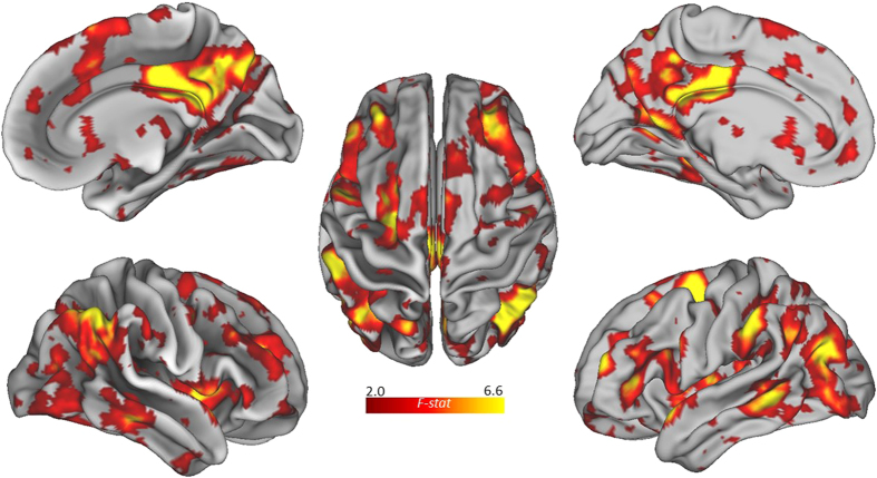
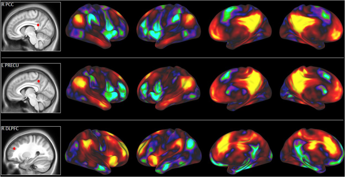
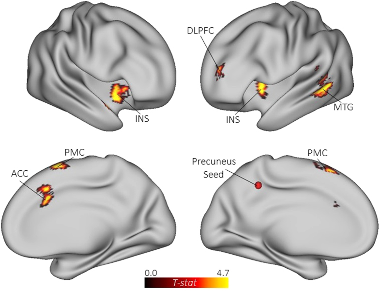
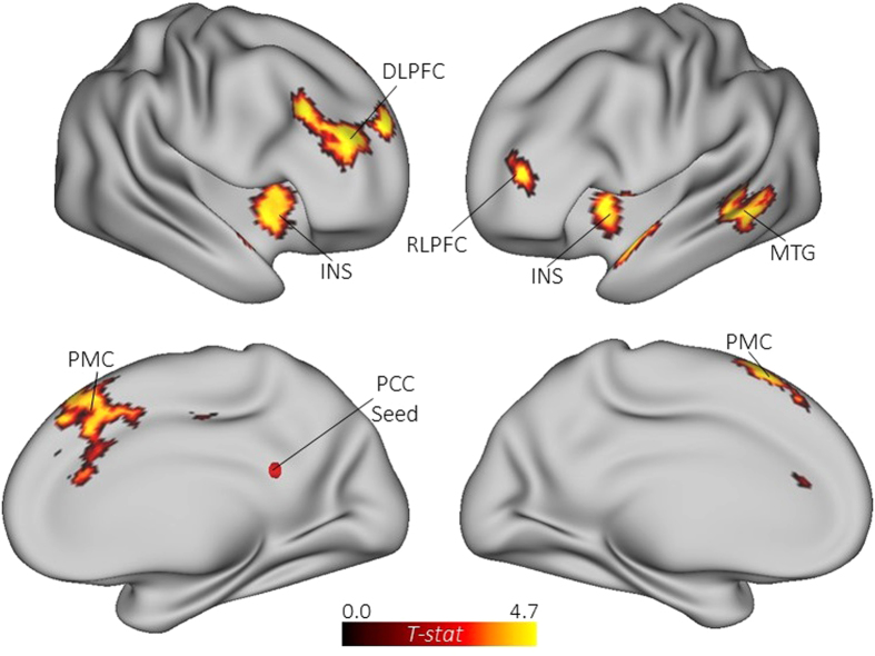
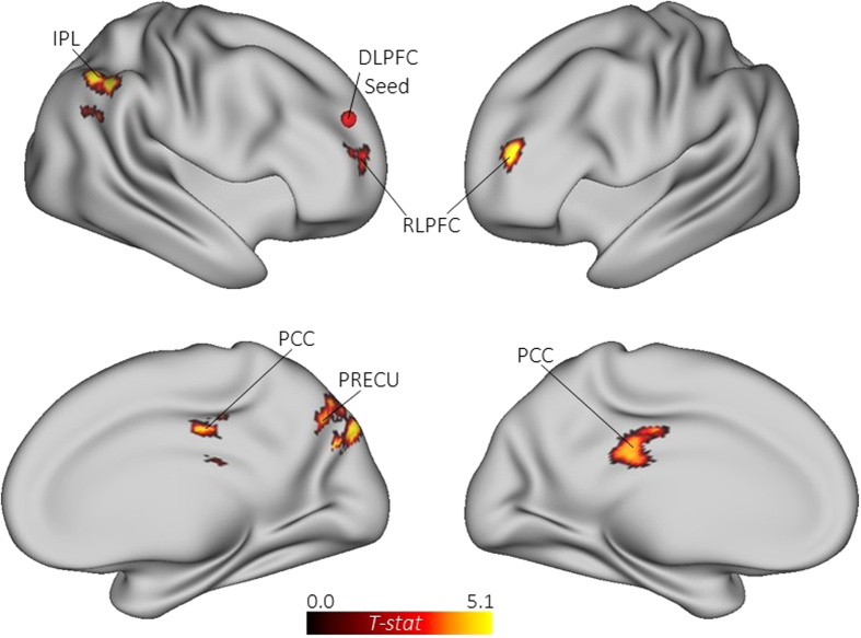
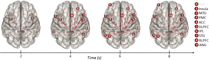
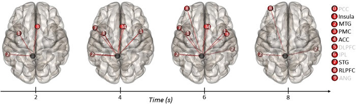
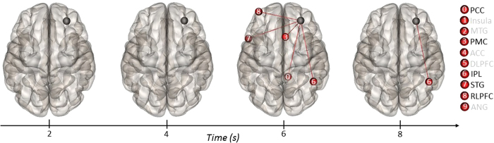
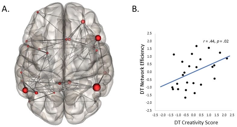
The role of attention in creative cognition remains controversial. Neuroimaging studies have reported activation of brain regions linked to both cognitive control and spontaneous imaginative processes, raising questions about how these regions interact to support creative thought. Using functional magnetic resonance imaging (fMRI), we explored this question by examining dynamic interactions between brain regions during a divergent thinking task. Multivariate pattern analysis revealed a distributed network associated with divergent thinking, including several core hubs of the default (posterior cingulate) and executive (dorsolateral prefrontal cortex) networks. The resting-state network affiliation of these regions was confirmed using data from an independent sample of participants. Graph theory analysis assessed global efficiency of the divergent thinking network, and network efficiency was found to increase as a function of individual differences in divergent thinking ability. Moreover, temporal connectivity analysis revealed increased coupling between default and salience network regions (bilateral insula) at the beginning of the task, followed by increased coupling between default and executive network regions at later stages. Such dynamic coupling suggests that divergent thinking involves cooperation between brain networks linked to cognitive control and spontaneous thought, which may reflect focused internal attention and the top-down control of spontaneous cognition during creative idea production.


Figures

Figure 1
Figure 1. Multivariate pattern analysis for the whole-brain task contrast (alternate uses > object characteristics).
Figure 2
Figure 2. Resting-state functional connectivity (RSFC) maps with select default and executive network ROIs.
Seeds were defined based on the whole-brain task contrast (alternate uses > object characteristics) and applied to an independent sample of participants (n = 42). Warm colors (red and yellow) reflect positive RSFC and cool colors (blue and green) reflect negative RSFC.
Figure 3
Figure 3. Functional connectivity maps for the general task contrast (alternate uses > object characteristics) with the left precuneus specified as a seed.
ACC = anterior cingulate cortex; DLPFC = dorsolateral prefrontal cortex; INS = insula; MTG = middle temporal gyrus; PMC = premotor cortex.
Figure 4
Figure 4. Functional connectivity maps for the general task contrast (alternate uses > object characteristics) with the right PCC specified as a seed.
DLPFC = dorsolateral prefrontal cortex; INS = insula; MTG = middle temporal gyrus; PCC = posterior cingulate cortex; PMC = premotor cortex; RLPFC = rostrolateral prefrontal cortex.
Figure 5
Figure 5. Functional connectivity maps for the general task contrast (alternate uses > object characteristics) with the right DLPFC specified as a seed.
DLPFC = dorsolateral prefrontal cortex; IPL = inferior parietal lobe; PCC = posterior cingulate cortex; PRECU = precuneus; RLPFC = rostrolateral prefrontal cortex.
Figure 6
Figure 6. ROI-to-ROI temporal connectivity for the general task contrast (alternate uses > object characteristics) with the right PCC specified as the source ROI (black sphere) and all other ROIs specified as targets (red spheres).
Regions labeled in black on the right show positive connectivity with the source ROI; regions labeled in gray were not significant.
Figure 7
Figure 7. ROI-to-ROI temporal connectivity for the general task contrast (alternate uses > object characteristics) with the left precuneus specified as the source ROI (black sphere) and all other ROIs specified as targets (red spheres).
Regions labeled in black on the right show positive connectivity with the source ROI; regions labeled in gray were not significant.
Figure 8
Figure 8. ROI-to-ROI temporal connectivity for general task contrast (alternate uses > object characteristics) with the right DLPFC specified as the source ROI (black sphere) and all other ROIs specified as targets (red spheres).
Regions labeled in black on the right show positive connectivity with the source ROI; regions labeled in gray were not significant.
Figure 9
Figure 9. Graph theory analysis of the functional network associated with divergent thinking.
(A) Nodes (ROIs from the whole-brain analysis) and edges (paths between the nodes) that were used to define the divergent thinking network. (B) Scatter plot depicting the correlation between composite creativity scores (i.e., average divergent thinking creativity ratings) and global efficiency of the divergent thinking network.

References

    1. Fox M. D. et al.. The human brain is intrinsically organized into dynamic, anticorrelated functional networks. Proc. Natl. Acad. Sci. USA, 102, 9673–9678 (2005). - PMC - PubMed
    1. Bressler S. & Menon V. Large-scale brain networks in cognition: Emerging methods and principles. Trends Cogn. Sci., 14, 277–290 (2010). - PubMed
    1. Spreng R. N., Sepulcre J., Turner G. R., Stevens W. D. & Schacter D. L. Intrinsic architecture underlying the relations among the default, dorsal attention, and frontoparietal control networks of the human brain. J. Cogn. Neurosci., 25, 74–86 (2013). - PMC - PubMed
    1. Sporns O. Contributions and challenges for network modes in cognitive neuroscience. Nat. Neurosci., 17, 652–660 (2014). - PubMed
    1. Gusnard D. A. & Raichle M. E. Searching for a baseline: Functional imaging and the resting human brain. Nat. Rev. Neurosci., 2, 685–694 (2001). - PubMed

LinkOut - more resources