Review
. 2015 Jul;67(3):601-55.
doi: 10.1124/pr.114.010249.

International Union of Basic and Clinical Pharmacology. XCVIII. Histamine Receptors

Affiliations
Review

International Union of Basic and Clinical Pharmacology. XCVIII. Histamine Receptors

Pertti Panula et al. Pharmacol Rev. 2015 Jul.

Abstract

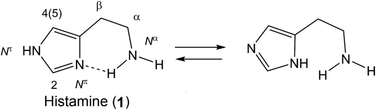
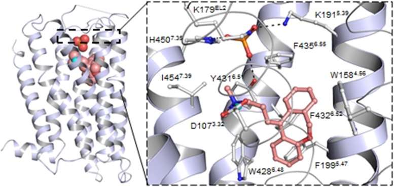
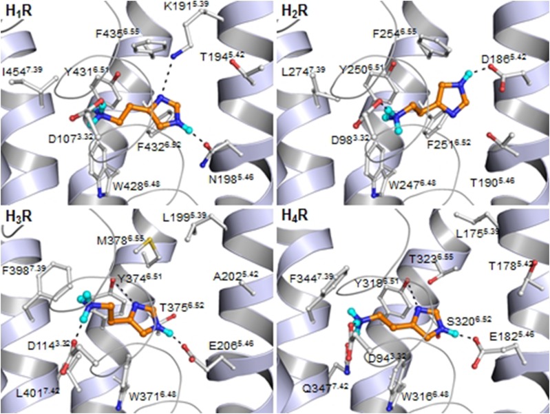
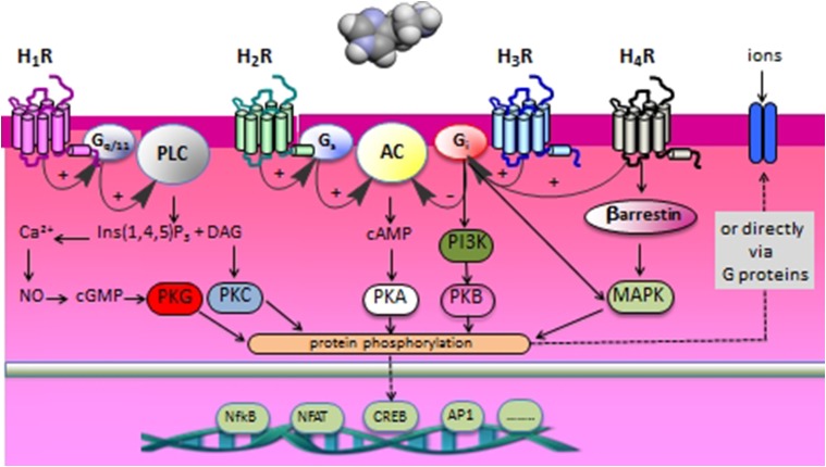
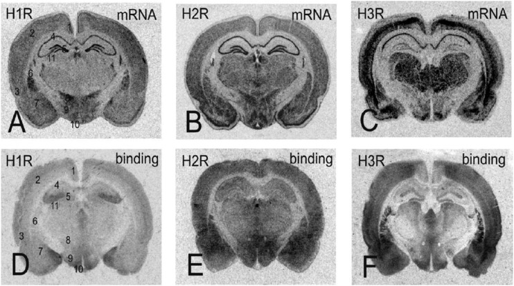
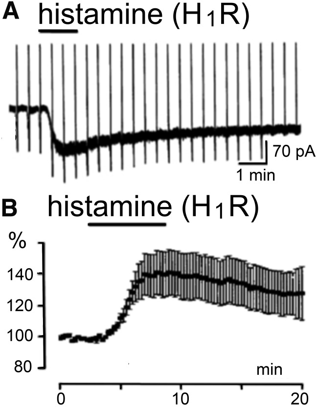
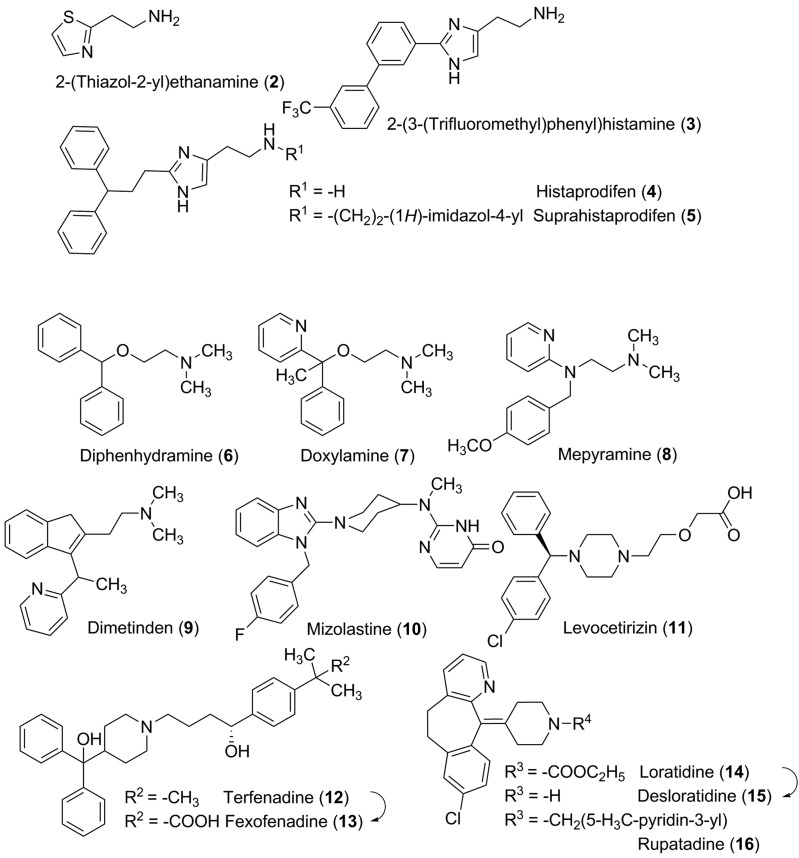
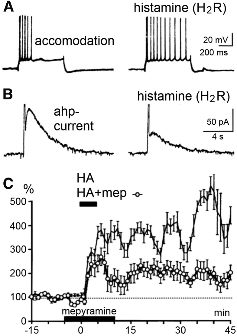
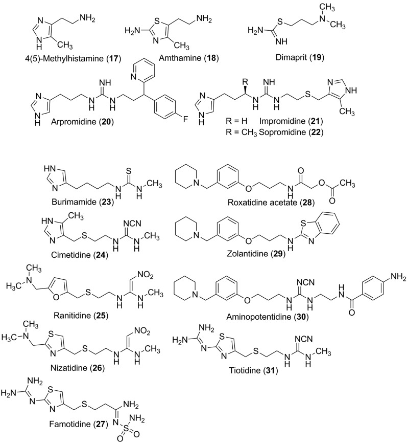
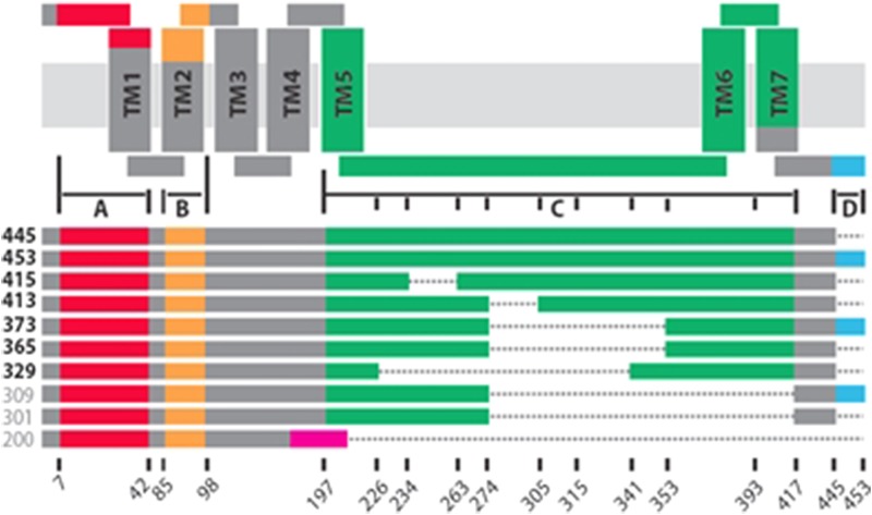
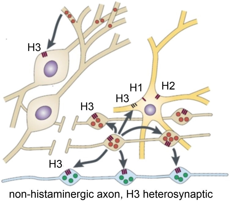
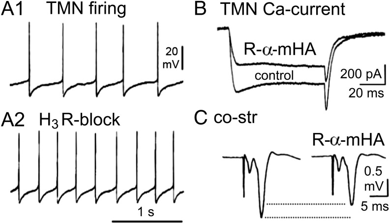
Histamine is a developmentally highly conserved autacoid found in most vertebrate tissues. Its physiological functions are mediated by four 7-transmembrane G protein-coupled receptors (H1R, H2R, H3R, H4R) that are all targets of pharmacological intervention. The receptors display molecular heterogeneity and constitutive activity. H1R antagonists are long known antiallergic and sedating drugs, whereas the H2R was identified in the 1970s and led to the development of H2R-antagonists that revolutionized stomach ulcer treatment. The crystal structure of ligand-bound H1R has rendered it possible to design new ligands with novel properties. The H3R is an autoreceptor and heteroreceptor providing negative feedback on histaminergic and inhibition on other neurons. A block of these actions promotes waking. The H4R occurs on immuncompetent cells and the development of anti-inflammatory drugs is anticipated.


Figures

Fig. 1.
Fig. 1.
Histamine.
Fig. 2.
Fig. 2.
Overall 7TM X-ray structure of the H1 receptor (PDB ID 3RZE) bound with the tricyclic H1 receptor inverse agonist doxepin and a phosphate ion and a close-up on the binding site with residues surrounding doxepin and the phosphate ion. Residues are numbered as found in the human H1 receptor sequence and with the corresponding Ballesteros-Weinstein numbering.
Fig. 3.
Fig. 3.
Binding of histamine in the four different histamine receptors as based on mutagenesis data and docking studies in the H1 receptor X-ray structure and H2 receptor–H4 receptor homology models, based on the H1 receptor X-ray structure. Residues are numbered as found in the human sequences and with the corresponding Ballesteros-Weinstein numbering.
Fig. 4.
Fig. 4.
Schematic overview of the main signal transduction routes of the four different histamine receptors.
Fig. 5.
Fig. 5.
Expression and receptor radioligand binding to cross sections of the rat brain. Data are shown for both mRNA distribution from in situ hybridization films and autoradiograms after labeling with radioactive ligands for each receptor. For identification of the brain areas, the following areas are marked in (A–D): 1, retrosplenial granular cortex; 2, primary somatosensory cortex; 3, entorhinal cortex; 4, CA1 area of the hippocampus; 5, dentate gyrus; 6, caudate putamen; 7, amygdala; 8, zona incerta; 9, lateral hypothalamic area; 10, arcuate nucleus; 11, dorsal lateral geniculate nucleus. Modified from Haas and Panula (2003).
Fig. 6.
Fig. 6.
Excitation of rat brainstem neurons by histamine through H1 receptor activation. (A) Inward current in a dorsal raphe neuron with increase in channel noise indicating (transient receptor potential channel-type) channel openings. (B) Increase in firing rate of 15 GABAergic (averaged) neurons in the substantia nigra (modified from Brown et al., 2002, and Korotkova et al., 2002).
Fig. 7.
Fig. 7.
H1 receptor ligands.
Fig. 8.
Fig. 8.
H2 receptor and cyclic AMP–mediated potentiation of excitation in rat hippocampal slices. (A) Block of the accommodation of firing in a CA1 pyramidal cell leading to a potentiation of the response to a depolarizing pulse. (B) Block of the IAHP current, which is responsible for a long-lasting Ca2+-dependent afterhyperpolarization and the accommodation of action potential firing. (C) Long-lasting potentiation of glutamatergic synaptic transmission (Schaffer collateral-CA1 pathway). The lower curve (open symbols) illustrates the pure H2 receptor mediated effect under mepyramine (8), the upper curve illustrates the boosting by H1 receptor signaling (which is sensitive to Li+). This long-lasting potentiation (several hours) has the characteristics of the protein kinase A–mediated LLTP (late phase of long-term potentiation) achieved here without high-frequency stimulation. H1 receptor activation alone causes no potentiation, rather an inhibitory effect presumably through activating a Ca2+-dependent K+-current (not shown here) (modified from Selbach et al., 1997).
Fig. 9.
Fig. 9.
H2 receptor ligands.
Fig. 10.
Fig. 10.
Schematic overview of several 7TM and truncated H3 receptor isoforms. Alternative splicing in regions A (red) and/or B (orange) results in partial deletion of the N-terminal domain and/or TM2 and EL1, respectively (isoforms not shown). Distinct splice events in region C (green) results in an IL3 of variable length and/or deletion of TM5, -6, and/or -7. Alternative splicing in region D (blue) adds 8 additional residues to the C-terminal tail of H3R. Finally, a frameshift after residue 171 results in a truncated receptor protein (magenta). Truncated H3 receptor isoforms are indicated in gray.
Fig. 11.
Fig. 11.
Neuronal targets of histaminergic neurons. H1 receptor and H2 receptor are located on many neurons where they modulate the activity in several ways (see text). H3 receptors are located on histaminergic neuron somata, dendrites, and axons to control firing, histamine synthesis, and release acting as autoreceptors. As heteroreceptors they control transmitter release from a wide variety of nonhistaminergic axons and in some cases inhibit the activity of nonhistaminergic neurons. Modified from Haas and Panula (2003).
Fig. 12.
Fig. 12.
Electrophysiological actions of H3 receptor activation in brain slices. (A1) Typical regular firing of a histaminergic tuberomamillary neuron. (A2) Block of the H3-autoreceptor activation by thioperamide releases the inhibition and thus increases firing rate. (B) Ca2+-current in response to depolarization of histaminergic neuron is reduced by the H3 receptor-agonist R-α-methylhistamine (33). (C) Glutamatergic cortico-striatal synaptic transmission (co-str) is reduced by R-α-methylhistamine, an example of an H3-heteroreceptor action.
Fig. 13.
Fig. 13.
H3 receptor agonists.
Fig. 14.
Fig. 14.
H3 receptor antagonists.
Fig. 15.
Fig. 15.
H4 receptor ligands.
Fig. 16.
Fig. 16.
Histamine gating chloride-channels. (A) Responses of artificially expressed GABAA receptor β3-homomeric channels in xenopus oocytes to GABA, tele-methylhistamine, histidine, and histamine all at 3 mM. (B) Thalamic GABAergic neuron is inhibited and hyperpolarized by histamine in a slice from ferret brain. An increased Cl-conductance (reversal potential −73 mV) displays H2 receptor pharmacology, see text. Modified from Lee et al. (2004) and Saras et al. (2008) with permission.
Fig. 17.
Fig. 17.
Ionic mechanism of histaminergic inhibitory postsynaptic potentials (ipsps) in aplysia. Paired recording in the histaminergic C2 neuron from aplysia and a follower neuron. Five action potentials are elicited (A) and the response is registered at different voltage levels (B, from −50 to −90 mV). Histamine is also puffed on the follower neuron at these different voltage levels (C). A fast component reverses at the equilibrium potential for Cl, between −60 and −70 mV, a slow component near −90 mV, close to the K+-equilibrium potential. Modified from McCaman and Weinreich (1985).
Fig. 18.
Fig. 18.
Histamine potentiates NMDA current. (A) Potentiation of NMDA-current in whole cell clamp with 1-second application of l-aspartate. (B) NMDA component (arrows) of excitatory postsynaptic potential (epsp) in hippocampal pyramidal cell. Modified from Vorobjev et al. (1993) and Yanovsky et al. (1995).

References

    1. Adami M, Pozzoli C, Menozzi A, Bertini S, Passeri B, Cantoni AM, Smits R, de Esch I, Leurs R, Coruzzi G. (2012) Effects of histamine H4 receptor ligands in a mouse model of gastric ulceration. Pharmacology 89:287–294. - PubMed
    1. Adjobo-Hermans MJ, Goedhart J, van Weeren L, Nijmeijer S, Manders EM, Offermanns S, Gadella TW., Jr (2011) Real-time visualization of heterotrimeric G protein Gq activation in living cells. BMC Biol 9:32. - PMC - PubMed
    1. Ahmad SF, Zoheir KM, Abdel-Hamied HE, Alrashidi I, Attia SM, Bakheet SA, Ashour AE, Abd-Allah AR. (2014) Role of a histamine 4 receptor as an anti-inflammatory target in carrageenan-induced pleurisy in mice. Immunology 142:374–383. - PMC - PubMed
    1. Aldi S, Takano K, Tomita K, Koda K, Chan NY-K, Marino A, Salazar-Rodriguez M, Thurmond RL, Levi R. (2014) Histamine H4-receptors inhibit mast cell renin release in ischemia/reperfusion via protein kinase C ε-dependent aldehyde dehydrogenase type-2 activation. J Pharmacol Exp Ther 349:508–517. - PMC - PubMed
    1. Alewijnse AE, Smit MJ, Hoffmann M, Verzijl D, Timmerman H, Leurs R. (1998) Constitutive activity and structural instability of the wild-type human H2 receptor. J Neurochem 71:799–807. - PubMed