. 2016 Jan;36(1):113-20.
doi: 10.1007/s10571-015-0225-3. Epub 2015 Jun 18.

Upregulation of miR-107 Inhibits Glioma Angiogenesis and VEGF Expression

Affiliations

Upregulation of miR-107 Inhibits Glioma Angiogenesis and VEGF Expression

Lei Chen et al. Cell Mol Neurobiol. 2016 Jan.

Erratum in

Abstract

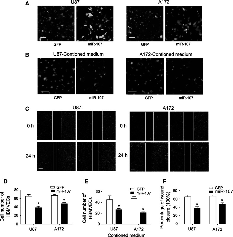
MicroRNAs can function as oncogenes or tumor suppressors in glioma. Previously, we showed that miR-107 inhibits glioma cell proliferation, migration, and invasion. Since tumor growth and invasion are closely related to angiogenesis, we further examined the role of miR-107 in glioma angiogenesis. In a co-culture of glioma cells and human brain microvascular endothelial cells (HBMVEC), overexpression of miR-107 in glioma cells led to the inhibition of HBMVEC proliferation, migration, and tube formation ability. ELISA, RT-PCR, and western blot assays revealed that upregulation of miR-107 in glioma cells inhibits VEGF expression. Our findings collectively support the critical involvement of miR-107 in glioma cell angiogenesis and highlight its potential as a therapeutic target for glioma.

Keywords: Angiogenesis; Glioma; HBMVEC; VEGF; miR-107.


Conflict of interest statement

No conflicts of interest were declared.

Figures

Fig. 1
Fig. 1
Upregulation of miR-107 inhibits glioma growth and angiogenesis in vivo. a Nude mice were subcutaneously inoculated with U87 cells transduced with GFP or miR-107, and neoplasms obtained 6 weeks later. Representative images are shown. b Comparison of tumor volumes between miR-107 and GFP groups every 7 days. c Quantification tumor weight after nude mice were subcutaneously inoculated with U87 cells transduced with GFP or miR-107. d Representative image of a neoplasm section stained for CD31 to determine vascular formation (×400 magnification). Scale bars 50 μm. e CD31-positive microvessels were counted in three different fields per section at ×400 magnification. *P < 0.05. GFP transduced with GFP only, miR-107 transduced with GFP and miR-107
Fig. 2
Fig. 2
Overexpression of miR-107 in glioma cells inhibits HBMVEC proliferation and migration in vitro. a U87 and A172 cells (green fluorescence, transduced with GFP or miR-107) were co-cultured with HBMVECs (red fluorescence, stained with PKH26) in six-well plates. Representative images, obtained 24 h later, are shown (×200 magnification). Scale bars 100 μm. b The effect of supernatant derived from U87 and A172 cells transduced with GFP or miR-107 on HBMVECs proliferation. Representative images are shown. Scale bars 100 μm. c Representative fluorescence microscopy image (×100 magnification) showing the effects of the supernatant fractions of U87 and A172 cells (transduced with GFP or miR-107) on HBMVEC (red fluorescence, stained with PKH26) migration in the wound-healing assay. Images were acquired directly after scratching (t = 0) and 24 h later (t = 24). Scale bars 200 μm. d The numbers of HBMVECs co-cultured with U87 and A172 glioma cells (transduced with GFP or miR-107) were calculated using ImageJ software. e Quantitation of the number of HBMVECs after treated with supernatant derived from U87 and A172 cells transduced with GFP or miR-107 using ImageJ software. f Quantitation of HBMVEC migration to the scratch using ImageJ software. *P < 0.05. GFP transduced with GFP only, miR-107 transduced with GFP and miR-107
Fig. 3
Fig. 3
Upregulation of miR-107 in glioma cells suppresses tubule formation in the HBMVEC and glioma cell co-culture system. a, b U87 or A172 glioma cells (green fluorescence, transduced with GFP or miR-107) and HBMVECs (red fluorescence, stained with PKH26) were co-cultured on Matrigel-coated plates, and tubule formation assessed after 48 h. Representative images are shown (×100). Scale bars 150 μm. c ImageJ software was applied to evaluate tubule crosses in a, b. *P < 0.05. GFP transduced with GFP only, miR-107 transduced with GFP and miR-107
Fig. 4
Fig. 4
Overexpression of miR-107 inhibits glioma VEGF expression. a ELISA assay data revealed that compared with the GFP group, VEGF expression levels are inhibited in the supernatant fractions of glioma cell lines, U87 and A172, transduced with miR-107. b RT-PCR disclosed downregulation of VEGF mRNA in U87 and A172 glioma cells transduced with miR-107. c Western blot results showed that miR-107 inhibits VEGF protein expression in U87 and A172 glioma cells. d Western blot assay showed that VEGF protein expression was downregulated in neoplasm treated with miR-107.*P < 0.05. GFP transduced with GFP only, miR-107 transduced with GFP and miR-107

References

    1. Ambros V (2004) The functions of animal microRNAs. Nature 431:350–355 - PubMed
    1. Bartel DP (2004) MicroRNAs: genomics, biogenesis, mechanism, and function. Cell 116:281–297 - PubMed
    1. Brandes AA, Franceschi E, Gorlia T, Wick W, Jacobs AH, Baumert BG, van den Bent M, Weller M, Stupp R (2012) Appropriate end-points for right results in the age of antiangiogenic agents: future options for phase II trials in patients with recurrent glioblastoma. Eur J Cancer 48:896–903 - PubMed
    1. Chen L, Chen XR, Zhang R, Li P, Liu Y, Yan K, Jiang XD (2013a) MicroRNA-107 inhibits glioma cell migration and invasion by modulating Notch2 expression. J Neurooncol 112:59–66 - PubMed
    1. Chen L, Zhang R, Li P, Liu Y, Qin K, Fa ZQ, Liu YJ, Ke YQ, Jiang XD (2013b) P53-induced microRNA-107 inhibits proliferation of glioma cells and down-regulates the expression of CDK6 and Notch-2. Neurosci Lett 534:327–332 - PubMed

Publication types

MeSH terms

Substances

LinkOut - more resources