. 2015 Aug;114(2):1286-97.
doi: 10.1152/jn.00010.2014. Epub 2015 Jun 17.

Single-motor-unit discharge characteristics in human lumbar multifidus muscle

Affiliations

Single-motor-unit discharge characteristics in human lumbar multifidus muscle

Lise R Lothe et al. J Neurophysiol. 2015 Aug.

Abstract

The underlying neurophysiology of postural control of the lower back in humans is poorly understood. We have characterized motor unit (MU) discharge activity in the deep lumbar multifidus (LM) muscle in nine healthy subjects (20-40 yr, 3 females). Bilateral fine wire electrodes were implanted at L4 spinal level using ultrasound guidance. EMG was recorded during spontaneous sitting and standing and during voluntary force production. Individual MUs were analyzed with regard to instantaneous discharge rate, interspike interval variability, alternation of activity between MUs, and cross correlation between concurrently active MUs quantified by the common drive coefficient (CDC). Significant effects of sitting vs. standing were seen on median discharge rate and interspike interval variability. Median discharge rate in 71 units was 5.4 and 6.9 pulses/s during spontaneous sitting and standing and 7.4 pulses/s during voluntary force production. Several MUs fired doublets. CDC analysis of 87 MU pairs showed a significantly higher common drive in spontaneous than in voluntary activity and significant differences between unilateral and bilateral pairs, although not when spontaneously active in standing. In spite of common drive, MUs were recruited from inactivity to tonic discharge lasting for several minutes without changes in discharge rate in already active MUs, and several instances were documented where activity was rotated between MUs. We argue that this behavior is indicative of self-sustained discharge in LM motoneurons, establishing intrinsic motoneuron properties as a central mechanism for postural control of deep back muscles.

Keywords: back; electromyography; motor activity; motor neurons; muscle tonus.


Figures

Fig. 1.
Fig. 1.
Bilateral raw fine-wire electromyography signals from deep lumbar multifidus (LM) during voluntary activation while standing. Instructions for movements are indicated below. The deep LM was eccentrically active in forward flexion and concentrically active in extension from flexion bilaterally, and unilaterally active in contralateral rotation and in ipsilateral hip extension. Additionally, in this subject, the right LM was active during contralateral hip extension. The flexion-relaxation phenomenon, where the LM was not active during full flexion (Floyd and Silver 1955; Watson et al. 1997; Colloca and Hinrichs 2005) is evident.
Fig. 2.
Fig. 2.
Unilateral recording showing concurrently active motor units (MUs) during 7 min of spontaneous standing (same subject as in Fig. 5). From bottom to top: unprocessed analog signal (black), discriminated MU action potentials (MUAPs) (color coded), superimposed smoothed discharge rates, and instantaneous discharge rates for individual units; pps, pulses/s. Insets, 3 top traces: superimposed consecutive MUAPs acquired over 10-s periods. The middle (blue) MUAPs remain similar in shape in spite of changes in amplitude. Changes in MUAP amplitude were interpreted to be caused by movements of the muscle fiber relative to the electrodes and often limited the duration of time periods where unequivocal MUAP sorting was possible. Note the synchrony in discharge rate fluctuations between concurrently active MUs indicating a common drive. In spite of this, there is rotation of activity among the MUs, probably reflecting activation and deactivation of self-sustained discharge in the individual motoneurons.
Fig. 3.
Fig. 3.
Bilateral recordings showing 5 concurrently active MUs (color coded) under voluntary forward flexion with auditory and visual feedback (same subject as in Fig. 6). A second-order high-pass Butterworth filter with cut-off frequency 0.75 Hz was applied to the smoothed discharge rate traces to remove mean frequency and low-frequency oscillations (top traces both sides) before common drive coefficient (CDC) analysis. Oscillations remain correlated (dashed lines) within and between sides even after removal of the larger respiratory related fluctuations seen in the single-unit instantaneous discharge rate traces, indicating a strong bilateral common drive.
Fig. 4.
Fig. 4.
Distribution of median MU discharge rates for spontaneous activity during sitting and for spontaneous and voluntary activity during standing. Boxes show medians and quartiles; whiskers denote 10 and 90 percentiles. Standing contributed significantly toward higher median discharge rates (see Table 1).
Fig. 5.
Fig. 5.
Unilateral recording from a single deep LM MU of a healthy, physically fit subject (same subject as in Fig. 2) during spontaneous standing. Slight changes in posture and breathing turned on and off MUAP trains all starting with 2 or 3 doublets with long postdoublet intervals (inset).
Fig. 6.
Fig. 6.
Unilateral recording from the deep LM during spontaneous standing, same subject as in Fig. 3. A, bottom to top: unprocessed analog signal (black), discriminated MUAPs (color coded), superimposed smoothed discharge rates, and instantaneous discharge rates for individual units. Top trace: repetitive doublet discharges occurring at low discharge rates. Insets: superimposed MUAPs during 10-s doublet discharge and 10-s nondoublet discharge. Note rotation of activity between the MUs. B: interspike interval histogram obtained during sustained doublet discharge. Note different time scales. C: Poincaré plot showing the relationship between 1,977 nondoublet interspike intervals (defined as interspike interval >20 ms) and their immediately following interspike intervals. Note that high-rate doublets are consistently preceded by low-rate intervals, and that the intradoublet instantaneous rate becomes lower as the instantaneous rate of the preceding interval increases (separate regression lines with 95% confidence interval for intervals followed by doublets and nondoublets).
Fig. 7.
Fig. 7.
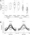
A: distribution of CDCs in 87 MU pairs with median recording duration of 30.5 s (range: 5.7-1,044 s). Spontaneous activity contributed significantly toward higher CDCs (see Table 1). B: cross correlograms from unilateral and bilateral recordings of MU pairs during spontaneous standing (cf. corresponding CDC values in A). CDC was defined as the maximum value within 0 ± 50 ms (hatched vertical lines).

References

    1. Bawa P, Calancie B. Repetitive doublets in human flexor carpi radialis muscle. J Physiol 339: 123–132, 1983. - PMC - PubMed
    1. Bawa P, Murnaghan C. Motor unit rotation in a variety of human muscles. J Neurophysiol 102: 2265–2272, 2009. - PubMed
    1. Bawa P, Pang MY, Olesen KA, Calancie B. Rotation of motoneurons during prolonged isometric contractions in humans. J Neurophysiol 96: 1135–1140, 2006. - PubMed
    1. Biering-Sørensen F. Physical measurements as risk indicators for low-back trouble over a one-year period. Spine 9: 106–119, 1984. - PubMed
    1. Burke RE, Rudomin P, Zajac FE. Catch property in single mammalian motor units. Science 168: 122–124, 1970. - PubMed

Publication types