. 2015 Aug 7;290(32):19629-40.
doi: 10.1074/jbc.M115.657874. Epub 2015 Jun 17.

Structural Basis for Antibody Recognition of Lipid A: INSIGHTS TO POLYSPECIFICITY TOWARD SINGLE-STRANDED DNA

Affiliations

Structural Basis for Antibody Recognition of Lipid A: INSIGHTS TO POLYSPECIFICITY TOWARD SINGLE-STRANDED DNA

Omid Haji-Ghassemi et al. J Biol Chem. .

Abstract

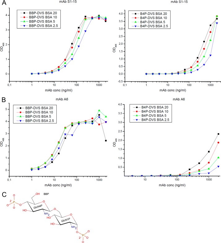
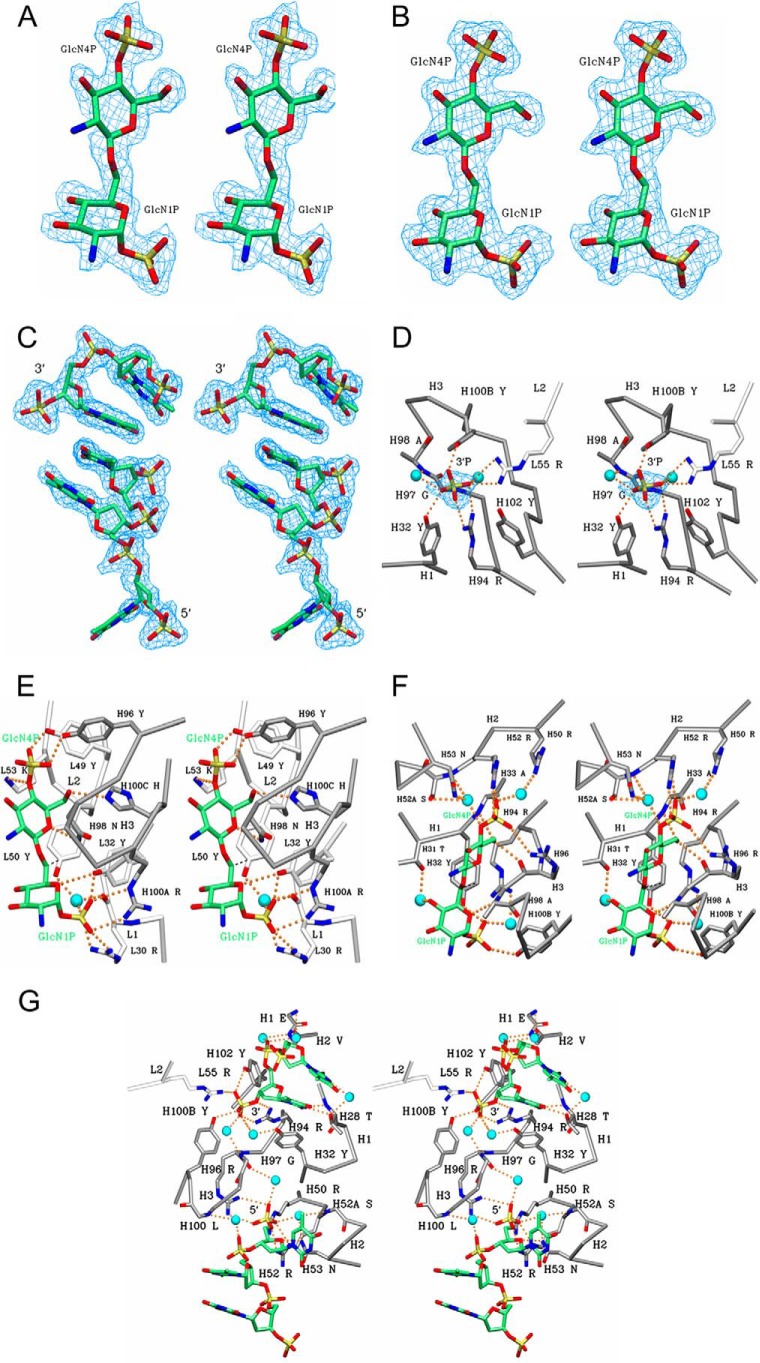
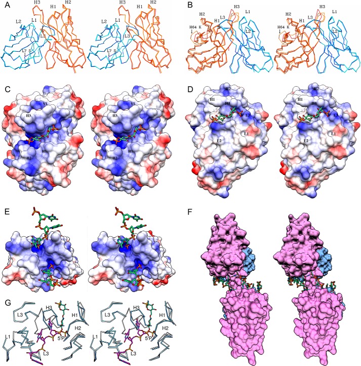
Septic shock is a leading cause of death, and it results from an inflammatory cascade triggered by the presence of microbial products in the blood. Certain LPS from Gram-negative bacteria are very potent inducers and are responsible for a high percentage of septic shock cases. Despite decades of research, mAbs specific for lipid A (the endotoxic principle of LPS) have not been successfully developed into a clinical treatment for sepsis. To understand the molecular basis for the observed inability to translate in vitro specificity for lipid A into clinical potential, the structures of antigen-binding fragments of mAbs S1-15 and A6 have been determined both in complex with lipid A carbohydrate backbone and in the unliganded form. The two antibodies have separate germ line origins that generate two markedly different combining-site pockets that are complementary both in shape and charge to the antigen. mAb A6 binds lipid A through both variable light and heavy chain residues, whereas S1-15 utilizes exclusively the variable heavy chain. Both antibodies bind lipid A such that the GlcN-O6 attachment point for the core oligosaccharide is buried in the combining site, which explains the lack of LPS recognition. Longstanding reports of polyspecificity of anti-lipid A antibodies toward single-stranded DNA combined with observed homology of S1-15 and A6 and the reports of several single-stranded DNA-specific mAbs prompted the determination of the structure of S1-15 in complex with single-stranded DNA fragments, which may provide clues about the genesis of autoimmune diseases such as systemic lupus erythematosus, thyroiditis, and rheumatic autoimmune diseases.

Keywords: autoimmunity; lipid A; lipopolysaccharide (LPS); monoclonal antibody; x-ray crystallography.


Figures

FIGURE 1.
FIGURE 1.
Quantitative conjugate ELISAs coated with graded concentrations of neoglycoconjugates corresponding to 2.5 (blue triangle), 5 (green triangle), 10 (red circle), and 20 (black square) pmol of ligand per well and reacted with mAbs (A) S1–15 and (B) A6 at concentrations indicated on the x axis are shown. Ligands used were bisphosphorylated lipid A backbone conjugated to BSA (BBP-divinyl sulfone BSA, left panel) and 4′-monophosphorylated lipid A backbone conjugated to BSA (right panel), and absorbance was measured at 405 nm. The chemical structure of the carbohydrate backbone of lipid A (BBP) used for crystallization trials (C). 1P and 4P refer to the position of the phosphates on the pyranose ring.
FIGURE 2.
FIGURE 2.
Stereo diagram 2FoFc electron density map (blue) contoured at 1. 0σ for the BBP lipid A analogue was observed in the combining sites of A6 (A) and S1–15 mAbs (B) and two single-stranded DNA fragments. p2(dT)p (top) and p3(dT)p (bottom) were observed in the combining site of S1–15 (C). To show the base stacking interaction, the trinucleotide (bottom) is bound to a symmetry-related Fab. Although the original antigen used was p5(dT)p, only two and three nucleotides could be modeled with confidence. Stereo diagram of S1–15 in complex with 5(dT)p ligand showing hydrogen bonds (orange dashed spheres) between residues of S1–15 and to the 3′P (D). The remainder of the antigen was disordered. Indirect hydrogen bonds mediated by waters (cyan spheres) are also shown. CDR loops of the light and heavy chain are colored white and gray, respectively. Color scheme is as follows: carbon, green; oxygen, red; nitrogen, blue; phosphorus, orange; water, cyan. Density (blue) for the phosphate is contoured at 1.0σ. Stereo view of A6 (E) and S1–15 (F) in complex with lipid A analogues BBP shows hydrogen bonds and water bridges between the antigen and S1–15. CDR loops of the light and heavy chain are colored white and gray, respectively. Strong hydrophobic contacts are shown as dashed lines (black). C6 hydroxyl group of second GlcN is the attachment point to inner core residues of LPS. Stereo diagram of S1–15 Fab in complex with two p5(dT)p ssDNA fragments is shown (G). Stereo diagram of S1–15 Fab in complex with two p5(dT)p ssDNA fragments is shown.
FIGURE 3.
FIGURE 3.
Stereo view of Fv structure alignments between liganded and unliganded structures of S1–15 (A) and A6 (B) is shown. Alignments were carried out using the α-carbon trace of the liganded VL as the reference structure for each antibody. Displacement of CDR L1 and H3 is highlighted. Dark blue, liganded light chain. Cyan, unliganded light chains. Orange, unliganded heavy chains. Red, liganded heavy chain. Because of conformational differences, both unliganded molecules in the asymmetric unit were included in the alignment of A6. Stereo images of the electrostatic surface potentials for Fv structures of S1–15 (C) and A6 (D), bound to lipid A analogue. Blue regions represent relatively positive surface charge, and red represents negative surface charge. White represents neutral surface charge. α-Carbon alignment between BV04-01 Fv (blue) and S1–15 Fv (white) in complex with a trinucleotide (khaki) and lipid A (green), respectively (E), is shown. Transparent surface diagram showing S1–15 Fv and symmetry-related Fv molecule in complex with p5(dT)p (green) fragments (F) is shown. The VL and VH domains are colored blue and pink, respectively. Stereo images of the electrostatic surface potentials for S1–15 Fv in complex with p5(dT)p ssDNA fragments. The lipid A 4′P binding pocket of S1–15 is closely related to the 5′P-binding site of BV04-01. For clarity, only the α-carbon trace of the CDRs is shown for each antibody (G).

References

    1. Angus D. C., Linde-Zwirble W. T., Lidicker J., Clermont G., Carcillo J., Pinsky M. R. (2001) Epidemiology of severe sepsis in the United States: Analysis of incidence, outcome, and associated costs of care. Crit. Care Med. 29, 1303–1310 - PubMed
    1. Engel C., Brunkhorst F. M., Bone H. G., Brunkhorst R., Gerlach H., Grond S., Gruendling M., Huhle G., Jaschinski U., John S., Mayer K., Oppert M., Olthoff D., Quintel M., Ragaller M., et al. (2007) Epidemiology of sepsis in Germany: results from a national prospective multicenter study. Intensive Care Med. 33, 606–618 - PubMed
    1. Martin G. S. (2012) Sepsis, severe sepsis and septic shock: changes in incidence, pathogens and outcomes. Expert Rev. Anti.-Infect. Ther. 10, 701–706 - PMC - PubMed
    1. Buttenschoen K., Radermacher P., Bracht H. (2010) Endotoxin elimination in sepsis: physiology and therapeutic application. Langenbeck. Arch. Surg. 395, 597–605 - PubMed
    1. Miller S. I., Ernst R. K., Bader M. W. (2005) LPS, TLR4 and infectious disease diversity. Nat. Rev. Microbiol. 3, 36–46 - PubMed

Publication types

MeSH terms

LinkOut - more resources