. 2015 Aug 14;290(33):20147-58.
doi: 10.1074/jbc.M115.655787. Epub 2015 Jun 17.

Porphyromonas gingivalis Stimulates Bone Resorption by Enhancing RANKL (Receptor Activator of NF-κB Ligand) through Activation of Toll-like Receptor 2 in Osteoblasts

Affiliations

Porphyromonas gingivalis Stimulates Bone Resorption by Enhancing RANKL (Receptor Activator of NF-κB Ligand) through Activation of Toll-like Receptor 2 in Osteoblasts

Ali Kassem et al. J Biol Chem. .

Abstract

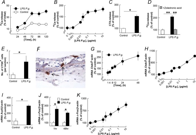
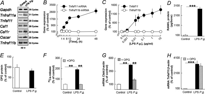
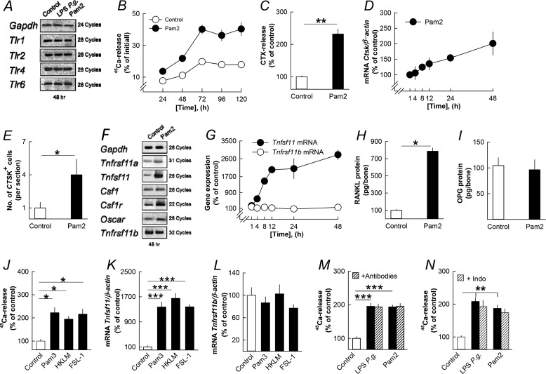
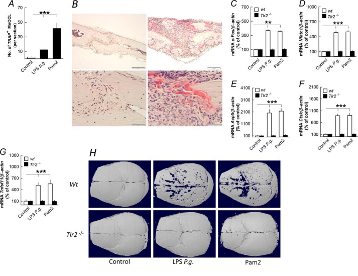
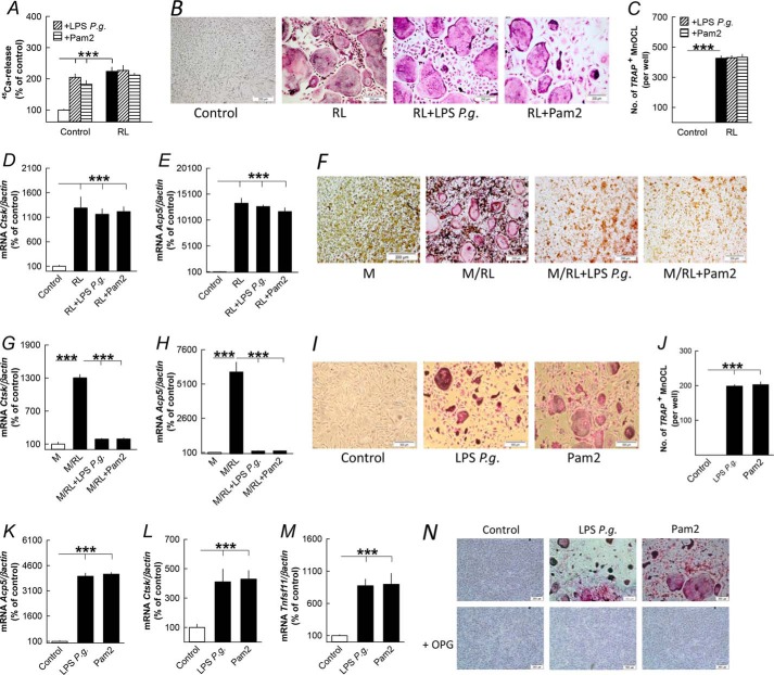
Periodontitis has been associated with rheumatoid arthritis. In experimental arthritis, concomitant periodontitis caused by oral infection with Porphyromonas gingivalis enhances articular bone loss. The aim of this study was to investigate how lipopolysaccharide (LPS) from P. gingivalis stimulates bone resorption. The effects by LPS P. gingivalis and four other TLR2 ligands on bone resorption, osteoclast formation, and gene expression in wild type and Tlr2-deficient mice were assessed in ex vivo cultures of mouse parietal bones and in an in vivo model in which TLR2 agonists were injected subcutaneously over the skull bones. LPS P. gingivalis stimulated mineral release and matrix degradation in the parietal bone organ cultures by increasing differentiation and formation of mature osteoclasts, a response dependent on increased RANKL (receptor activator of NF-κB ligand). LPS P. gingivalis stimulated RANKL in parietal osteoblasts dependent on the presence of TLR2 and through a MyD88 and NF-κB-mediated mechanism. Similarly, the TLR2 agonists HKLM, FSL1, Pam2, and Pam3 stimulated RANKL in osteoblasts and parietal bone resorption. LPS P. gingivalis and Pam2 robustly enhanced osteoclast formation in periosteal/endosteal cell cultures by increasing RANKL. LPS P. gingivalis and Pam2 also up-regulated RANKL and osteoclastic genes in vivo, resulting in an increased number of periosteal osteoclasts and immense bone loss in wild type mice but not in Tlr2-deficient mice. These data demonstrate that LPS P. gingivalis stimulates periosteal osteoclast formation and bone resorption by stimulating RANKL in osteoblasts via TLR2. This effect might be important for periodontal bone loss and for the enhanced bone loss seen in rheumatoid arthritis patients with concomitant periodontal disease.

Keywords: bone; inflammation; innate immunity; osteoblast; osteoclast; toll-like receptor (TLR).


Figures

FIGURE 1.
FIGURE 1.
LPS from P. gingivalis (P.g.) stimulates bone resorption, osteoclast formation, and expression of osteoclastic and osteoclastogenic genes in organ cultures of neonatal mouse parietal bones. A–C, LPS P. gingivalis time- and concentration-dependently increased 45Ca and CTX release from the parietal bones. D, the stimulatory effect by LPS P. gingivalis on 45Ca release was inhibited by zoledronic acid (0.2 μmol/liter). E, the number of cathepsin K-positive (CTSK+) osteoclasts was enhanced by LPS P. gingivalis. G and H, LPS P. gingivalis time- and concentration-dependently enhanced the mRNA expression of Ctsk. I and J, the mRNA expression of Acp5 (48 h) and c-fos (1 and 48 h) was increased by LPS P. gingivalis. K, LPS P. gingivalis concentration-dependently increased c-fos mRNA. *, p < 0.05; **, p < 0.01 compared with unstimulated controls (C–E, I, and J) or to LPS P. gingivalis-stimulated bones (D). LPS P. gingivalis was used at a concentration of 10 μg/ml in A, C–G, I, and J. Data are the means of 4–5 observations, and S.E. is given as vertical bars when larger than the radius of the symbol.
FIGURE 2.
FIGURE 2.
The stimulatory effect on bone resorption in neonatal mouse parietal bones by LPS from P. gingivalis (P.g.) is dependent on increased RANKL. A, LPS P. gingivalis enhanced the mRNA expression of Tnfrsf11a, Tnfsf11, Csf1, Csf1r, and Oscar without affecting Tnfrsf11b. B and C, LPS P. gingivalis time- and concentration-dependently enhanced Tnfsf11 mRNA with no effect on Tnfrsf11b mRNA. D and E, LPS P. gingivalis enhanced the cellular level of RANKL protein without affecting OPG protein. F–H, the stimulatory effect by LPS P. gingivalis on 45Ca release, and Ctsk mRNA was inhibited by adding exogenous OPG (300 ng/ml) to the culture medium, whereas Tnfsf11 mRNA was unaffected. **, p < 0.01; ***, p < 0.001 compared with unstimulated controls (D and F–H) or to LPS P. gingivalis-stimulated bones (F and G). LPS P. gingivalis was used at a concentration of 10 μg/ml in A, B, and D–H. Data are the means of 4–5 observations, and S.E. is given as vertical bars when larger than the radius of the symbol.
FIGURE 3.
FIGURE 3.
The lipopeptide Pam2 and three additional TLR2 agonists stimulate bone resorption, osteoclast formation, and expression of osteoclastic and osteoclastogenic genes in organ cultures of neonatal mouse parietal bones by an effect dependent on RANKL but independent on cytokine and prostaglandin formation. A, LPS P. gingivalis and Pam2 did not affect the mRNA expression of Tlr1, Tlr2, Tlr4, and Tlr6. B–E, Pam2 increased the release of 45Ca (B) and CTX (C), up-regulated Ctsk mRNA (D), and enhanced the number of cathepsin K positive (CTSK+) osteoclasts (E). F, Pam2 increased the mRNA expression of Tnfrsf11a, Tnfsf11, Csf1, Csf1r, and Oscar without affecting Tnfrsf11b. G–I, Pam2 time-dependently increased Tnfsf11 mRNA (G) resulting in increased RANKL protein after 48 h (H) without affecting Tnfrsf11b mRNA (G) or OPG protein (I). J–L, Pam3 (10 ng/ml), HKLM (107 colony-forming units (CFU)) and FSL-1 (0.1 μg/ml)-stimulated 45Ca release (J) and the mRNA expression of Tnfsf11 (K) without affecting Tnfrsf11b (L). M and N, the stimulatory effect by LPS P. gingivalis and Pam2 on 45Ca release was unaffected by adding a mixture of antibodies neutralizing IL-1β, IL-6, IL-11, LIF, OSM, and TNF-α or by adding indomethacin (1 μmol/liter). *, p < 0.05; **, p < 0.01; ***, p < 0.001 compared with unstimulated controls (C, E, H, J, K, M, and N). LPS P. gingivalis (P.g.) was used at a concentration of 10 μg/ml in A, M, and N. Pam2 was used at a concentration of 10 ng/ml in A–I, M, and N. Data are the means of four-five observations, and S.E. is given as vertical bars when larger than the radius of the symbol.
FIGURE 4.
FIGURE 4.
Five different TLR2 agonists enhanced Tnfsf11 mRNA expression in mouse parietal osteoblasts by a mechanism dependent on TLR2, MyD88, and NF-κB but independent on cytokine formation. A, LPS P. gingivalis (P.g.) and Pam2 up-regulated Tlr2 without affecting Tlr1, Tlr4 or Tlr6. B and C, LPS P. gingivalis time- and concentration-dependently enhanced Tnfsf11 mRNA without affecting Tnfrsf11b. D, Pam2 time-dependently increased Tnfsf11 mRNA without affecting Tnfrsf11b. E, Pam3 (10 ng/ml), HKLM (107 CFU), and FSL-1 (0.1 μg/ml) stimulated Tnfsf11 mRNA. F–H, the stimulatory effect by LPS P. gingivalis and Pam2 on Tnfsf11 mRNA was unaffected by adding antibodies neutralizing IL-1β, IL-6, IL-11, LIF, OSM, and TNF-α. I, LPS P. gingivalis, Pam2, Pam3 (10 ng/ml), HKLM (107 CFU), and FSL-1 (0.1 μg/ml), but not LPS from E. coli (10 μg/ml), increased Tnfsf11 mRNA in osteoblasts from wild type (Wt) but not from Tlr2-deficient mice. J, LPS P. gingivalis, Pam2, Pam3 (10 ng/ml), HKLM (107 CFU), and FSL-1 (0.1 μg/ml) enhanced Tnfsf11 mRNA in osteoblasts from wild-type mice but not from MyD88-deficient mice. K, LPS P. gingivalis and Pam2 increased the mRNA expression of the four NF-κB subunits p50, p52, p65, and RelB. L, LPS P. gingivalis and Pam2 increased NF-κB-driven luciferase in transfected osteoblasts. M and N, the stimulatory effect by LPS P. gingivalis and Pam2 on Tnfsf11 (M) and Il6 mRNA (N) was inhibited by the two NF-κB inhibitors BMS (10 μmol/liter) and Celastrol (0.2 μmol/liter). **, p < 0.01; ***, p < 0.001 compared with unstimulated controls (E–K) or to LPS P. gingivalis-stimulated osteoblasts (M and N). LPS P. gingivalis was used at a concentration of 10 μg/ml in A, B, and F–K. Pam2 was used at a concentration of 10 ng/ml in A, D, and F–N. Data are the means of four-five observations, and S.E. is given as vertical bars when larger than the radius of the symbol.
FIGURE 5.
FIGURE 5.
Injection of LPS from P. gingivalis (P.g.) and the TLR2 agonist Pam2 above skull bones stimulates osteoclast formation, expression of osteoclastic and osteoclastogenic genes, and bone loss in skull bones from 5-week-old mice. A and B, LPS P. gingivalis and Pam2 enhanced the number of tartrate-resistant acid phosphatase-positive, multinucleated osteoclasts (TRAP+MuOCL); the left panel of B shows a parietal bone 6 days after injection of vehicle, and the right panel of B shows osteoclasts in parietal bones 6 days after injection of LPS P. gingivalis. C–G, injection of LPS P. gingivalis or Pam2 increased the mRNA expression of c-fos, Nfatc1, Acp5, Ctsk, and Tnfsf11 after 3 days in skull bones from wild type (wt) but not from Tlr2-deficient mice. H, injection of LPS P. gingivalis or Pam2 resulted in bone loss after 6 days in skull bones from wild type but not in Tlr2-deficient mice. Images shown are representative of seven images per group. **, p < 0.01; ***, p < 0.001 compared with unstimulated controls (A and C–G). Data are the means of six-seven observations, and S.E. is given as vertical bars.
FIGURE 6.
FIGURE 6.
LPS from P. gingivalis and the TLR2 agonist Pam2 regulates bone resorption and osteoclast formation differently in parietal bones, periosteal/endosteal bone cell, and bone marrow cell cultures primed by RANKL (RL). A, RANKL (10 ng/ml)-stimulated 45Ca release from neonatal mouse parietal bones in organ culture was not affected by LPS P. gingivalis (10 μg/ml) or Pam2 (10 ng/ml). B–E, RL (10 ng/ml) stimulation of tartrate-resistant acid phosphatase-positive, multinucleated osteoclasts (TRAP+MuOCL), and mRNA expression of Ctsk and Acp5 in periosteal/endosteal cell cultures from mouse parietal bone were not affected by co-treatment with LPS P. gingivalis (P.g., 10 μg/ml) or Pam2 (10 ng/ml). F–H, increased formation of TRAP+ multinucleated osteoclasts and mRNA expression of Ctsk and Acp5 in M-CSF (30 ng/ml)- and RL (4 ng/ml)-stimulated mouse bone marrow cell cultures were abolished by co-treatment with LPS P. gingivalis and Pam2. I–M, LPS P. gingivalis (10 μg/ml) and Pam2 (10 ng/ml) increased formation of TRAP+ multinucleated osteoclasts and mRNA expression of Acp5, Ctsk and Tnfsf11 in periosteal/endosteal cell cultures from mouse parietal bone. N, the stimulatory effect by LPS P. gingivalis and Pam2 on osteoclast formation in periosteal/endosteal cell cultures was abolished by adding OPG (300 ng/ml) to the culture medium. ***, p < 0.001 compared with unstimulated controls. Data are the means of six-seven observations, and S.E. is given as vertical bars.

References

    1. Walsh N. C., Gravallese E. M. (2010) Bone remodeling in rheumatic disease: a question of balance. Immunol. Rev. 233, 301–312 - PubMed
    1. Bartold P. M., Cantley M. D., Haynes D. R. (2010) Mechanisms and control of pathologic bone loss in periodontitis. Periodontol. 2000 53, 55–69 - PubMed
    1. Schett G., Gravallese E. (2012) Bone erosion in rheumatoid arthritis: mechanisms, diagnosis and treatment. Nat. Rev. Rheumatol. 8, 656–664 - PMC - PubMed
    1. Souza P. P., Lerner U. H. (2013) The role of cytokines in inflammatory bone loss. Immunol Invest. 42, 555–622 - PubMed
    1. Lorenzo J., Horowitz M., Choi Y. (2008) Osteoimmunology: interactions of the bone and immune system. Endocr. Rev. 29, 403–440 - PMC - PubMed

Publication types

LinkOut - more resources