. 2015 Aug 21;290(34):20804-20814.
doi: 10.1074/jbc.M115.669440. Epub 2015 Jun 17.

Mia40 Protein Serves as an Electron Sink in the Mia40-Erv1 Import Pathway

Affiliations

Mia40 Protein Serves as an Electron Sink in the Mia40-Erv1 Import Pathway

Sonya E Neal et al. J Biol Chem. .

Abstract

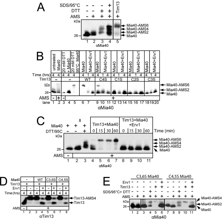
A redox-regulated import pathway consisting of Mia40 and Erv1 mediates the import of cysteine-rich proteins into the mitochondrial intermembrane space. Mia40 is the oxidoreductase that inserts two disulfide bonds into the substrate simultaneously. However, Mia40 has one redox-active cysteine pair, resulting in ambiguity about how Mia40 accepts numerous electrons during substrate oxidation. In this study, we have addressed the oxidation of Tim13 in vitro and in organello. Reductants such as glutathione and ascorbate inhibited both the oxidation of the substrate Tim13 in vitro and the import of Tim13 and Cmc1 into isolated mitochondria. In addition, a ternary complex consisting of Erv1, Mia40, and substrate, linked by disulfide bonds, was not detected in vitro. Instead, Mia40 accepted six electrons from substrates, and this fully reduced Mia40 was sensitive to protease, indicative of conformational changes in the structure. Mia40 in mitochondria from the erv1-101 mutant was also trapped in a completely reduced state, demonstrating that Mia40 can accept up to six electrons as substrates are imported. Therefore, these studies support that Mia40 functions as an electron sink to facilitate the insertion of two disulfide bonds into substrates.

Keywords: disulfide; mitochondria; oxidation-reduction (redox); protein import; redox; redox regulation; thiol.


Figures

FIGURE 1.
FIGURE 1.
Treatment with reductant decreases the rate of Tim13 oxidation by Mia40 and Erv1. A, gel system was calibrated with Tim13 mutants in which the 1st and 4th cysteines (C1,4S) and the 2nd and 3rd cysteines (C2,3S) have been mutated to serine residues. Reduced Tim13 and mutants were left untreated (lanes 1–3) or treated with AMS (lanes 4–6). B, reduced Tim13 (15 μm) was incubated with combinations of Mia40 (1 μm) and Erv1 (1 μm) in a time course assay as indicated (lanes 5–13). Aliquots were removed, and the free thiols were blocked with 20 mm AMS treatment. Oxidized (Tim13) and reduced (Tim13-AMS4) Tim13 were detected by nonreducing SDS-PAGE followed by immunoblotting with antibodies against Tim13. To calibrate the gel system, WT Tim13 and Tim13 with the 2nd and 4th cysteines mutated to serine residues (Tim13 C2,4S) were included. Reduced Tim13 samples were left untreated (lanes 1 and 3) or treated with AMS (lanes 2 and 4). The asterisk denotes nonspecific bands (lanes 2 and 4). C, as indicated in B with the addition of 5 mm (lanes 7–10) and 10 mm (lanes 11–14) GSH. D, as indicated in B with the addition of 5 mm ascorbate in the oxidation reaction. E, reactions in the presence of 5 mm GSH and 5 mm ascorbate were quantitated using a Bio-Rad FX Molecular Imager and the affiliated Quantity 1 software; 100% was set as the amount of Tim13 that was oxidized at the end point in the absence of reductant (n = 3). Results with 10 mm GSH were identical to those of ascorbate and are not shown to simplify the figure.
FIGURE 2.
FIGURE 2.
Reductant treatment decreases in organello import of Cmc1 and Tim13, but not TIM23 substrate, Su9-DHFR. A, mitochondria were preincubated with 2 or 5 mm GSH for 10 min followed by the import of Tim13 or Cmc1. Protease treatment of mitochondria removed the precursor that failed to import, and the imported precursors were analyzed by reducing SDS-PAGE and autoradiography. A 10% standard (Std) from the translation reaction was included. B, as indicated in A but with ascorbate. C, as in A, except Su9-DHFR was imported in the presence of GSH. D, as in B, except Su9-DHFR was imported in the presence of ascorbate. Import reactions were quantitated, and 100% was set as the amount of precursor imported into WT mitochondria at the end point in the time course. Representative gels are shown (n = 4).
FIGURE 3.
FIGURE 3.
Mixed disulfide analysis shows that Mia40 is reduced by Tim13 in vitro. A, reduced Tim13 was preincubated in the absence or presence (+ IAA pretreatment of Tim13, lanes 3, 5, 7, 11, 13, 17, and 19) of IAA as indicated. Subsequently, equimolar amounts of recombinant Mia40, Erv1, and Tim13 were incubated in combination as indicated for 10 min. All samples were incubated with IAA post-treatment to block free thiols. Samples were separated by nonreducing SDS-PAGE followed by immunoblot analysis with antibodies against Tim13 (lanes 1–7), Mia40 (lanes 8–13), and Erv1 (lanes 14–19). Asterisks denote multimers of Tim13; the diamond marks a Mia40 dimer, and the square marks an Erv1 dimer (19). B, as in A, samples were separated on a reducing gel. Representative gels are shown (n = 4).
FIGURE 4.
FIGURE 4.
Mia40 can accept up to six electrons from Tim13. A, control reactions to assess the Mia40 oxidation status. 1 μm Mia40 was left untreated (lane 1), treated with 20 mm AMS for 60 min (lane 2), incubated in 20 mm DTT for 60 min followed by 20 mm AMS addition (lane 3), or incubated at 95 °C with 0.05% SDS for 10 min followed by incubation in 20 mm DTT for 60 min and subsequent 20 mm AMS addition (lane 4). 15 μm reduced Tim13 was incubated with oxidized Mia40 at 25 °C for 60 min followed by addition of 20 mm AMS (lane 5). Proteins were separated by nonreducing SDS-PAGE and Mia40 was detected by immunoblot analysis. B, redox state of Mia40 was determined by incubation of 15 μm reduced Tim13 (WT, or C1S, C2S, C3S, and C4S mutants) with either 1 μm Mia40 or 1 μm Mia40 and 1 μm Erv1 for up to 4 h followed by 20 mm AMS addition (lanes 6–20). The samples were resolved by nonreducing SDS-PAGE and immunoblotted with anti-Mia40. In control reactions, Mia40 was left untreated for 4 h (lanes 1 and 2), reduced with 20 mm DTT (lanes 3 and 4), and heated to 95 °C with addition of 0.05% SDS (lane 5) followed by AMS treatment as indicated. C, as in B except reduced Tim13 (15 μm) was incubated with combinations of 1 μm Mia40 or 1 μm Mia40 and 1 μm Erv1 in a time course assay as indicated (lanes 4–11). Aliquots were removed at the indicated times and AMS was added. In control reactions, Mia40 was incubated in reaction buffer for 60 min (lanes 1 and 2) or in the presence of 20 mm DTT and 95 °C (lane 3) followed by 20 mm AMS addition (lanes 2 and 3). D, reduced Tim13 (15 μm) was incubated with a combination of WT Mia40 or Mia40 mutants C3,6S and C4,5S (1 μm) and Erv1 (1 μm) for 4 h. The reaction was stopped, and free thiols were blocked with AMS treatment. Oxidized (Tim13) and reduced (Tim13-AMS4) Tim13 were separated on nonreducing SDS-PAGE followed by immunoblotting with antibodies against Tim13. E, as in D, except that the redox status of mutant Mia40 (C3,6S and C4,5S) was investigated by treatment of AMS and subsequent nonreducing SDS-PAGE and immunoblot analysis with anti-Mia40 (n = 3).
FIGURE 5.
FIGURE 5.
Mia40 is reduced by Tim13 at an equimolar cysteine concentration. The redox state of WT Mia40 (A), C3,6S Mia40 (B), or C4,5S Mia40 (C) was determined by incubating increasing amounts of reduced Tim13 with 1 μm Mia40 for 2 h followed by addition of 20 mm AMS. The protein ratio (μm Tim13:μm Mia40) and cysteine ratio (Tim13 cysteines/Mia40 cysteines) are indicated. In control reactions, Mia40 was reduced with 20 mm DTT and incubated at 25 or 95 °C in the presence of 0.05% SDS along with 20 mm DTT. A representative gel is shown (n = 3).
FIGURE 6.
FIGURE 6.
Tim13 induces conformational changes in Mia40. A, reduced Tim13 (15 μm) was incubated with Mia40 (1 μm) followed by addition of 5 μg/ml trypsin at 25 °C. At the indicated times, aliquots were removed, and soybean trypsin inhibitor was added. Control reactions include untreated Mia40 and Mia40 incubated in 20 mm DTT at 95 °C. Samples were separated by SDS-PAGE followed by immunoblot analysis with antibodies against Mia40. A representative gel is shown. B, as in A, in the presence of 15 μm BSA (n = 4).
FIGURE 7.
FIGURE 7.
Mia40 is reduced in the erv1–101 mutant at the restrictive temperature 37 °C. A, redox status of Mia40 was analyzed in the presence of WT and Erv1 mutants (C30S or C133S) (lanes 6–8). Reduced Tim13 (15 μm) was incubated with combinations of Mia40 (1 μm), Erv1 (1 μm), and mutant Erv1 (C133S or C30S, 1 μm). Aliquots were removed at 4 h, and free thiols were blocked with AMS. Mia40 was separated by nonreducing SDS-PAGE followed by immunoblotting with antibodies against Mia40. Control reactions for Mia40 were included as described previously in Fig. 4A (lanes 1–4). B, reduced Cmc1 (15 μm) was incubated in combination with Mia40 (1 μm) and/or Erv1 (1 μm) for 4 h, followed by AMS treatment (lanes 5 and 6). Samples were separated by nonreducing SDS-PAGE followed by immunoblot analysis with antibodies against Mia40. Arrows depict oxidized Mia40, reduced Mia40 (Mia40-AMS6), a Cmc1-Mia40 complex, and the Mia40 dimer. Control reactions for Mia40 (lanes 1–4) as described in A. C, radiolabeled WT Mia40 and Mia40 CPC-SX9S-SX9S (mutant that lacks the four cysteines that form the structural disulfide bonds) were synthesized in vitro in a reticulocyte transcription/translation system. Aliquots were removed at 90 min, and free thiols on Mia40 were blocked with AMS and separated by nonreducing SDS-PAGE. D, redox state of Mia40 was investigated in mitochondria isolated from WT and erv1-101 yeast strains grown at the permissive temperature (25 °C) or nonpermissive temperature (37 °C). Isolated mitochondria were precipitated in 20% TCA in the presence of AMS. In control reactions, WT mitochondria were left untreated or incubated with 20 mm DTT at 25 or 95 °C prior to TCA precipitation and AMS treatment. Proteins were resolved on nonreducing SDS-PAGE and immunoblotted with anti-Mia40. Representative gels are shown (n = 4).

References

    1. Chacinska A., Koehler C. M., Milenkovic D., Lithgow T., Pfanner N. (2009) Importing mitochondrial proteins: machineries and mechanisms. Cell 138, 628–644 - PMC - PubMed
    1. Herrmann J. M., Riemer J. (2012) Mitochondrial disulfide relay: redox-regulated protein import into the intermembrane space. J. Biol. Chem. 287, 4426–4433 - PMC - PubMed
    1. Koehler C. M., Tienson H. L. (2009) Redox regulation of protein folding in the mitochondrial intermembrane space. Biochim. Biophys. Acta 1793, 139–145 - PMC - PubMed
    1. Stojanovski D., Bragoszewski P., Chacinska A. (2012) The MIA pathway: a tight bond between protein transport and oxidative folding in mitochondria. Biochim. Biophys. Acta 1823, 1142–1150 - PubMed
    1. Koehler C. M., Merchant S., Schatz G. (1999) How membrane proteins travel across the mitochondrial intermembrane space. Trends Biochem. Sci. 24, 428–432 - PubMed

Publication types

MeSH terms

Substances