. 2015 Sep;89(17):8840-54.
doi: 10.1128/JVI.01232-15. Epub 2015 Jun 17.

Cocrystal Structures of Antibody N60-i3 and Antibody JR4 in Complex with gp120 Define More Cluster A Epitopes Involved in Effective Antibody-Dependent Effector Function against HIV-1

Affiliations

Cocrystal Structures of Antibody N60-i3 and Antibody JR4 in Complex with gp120 Define More Cluster A Epitopes Involved in Effective Antibody-Dependent Effector Function against HIV-1

Neelakshi Gohain et al. J Virol. 2015 Sep.

Abstract

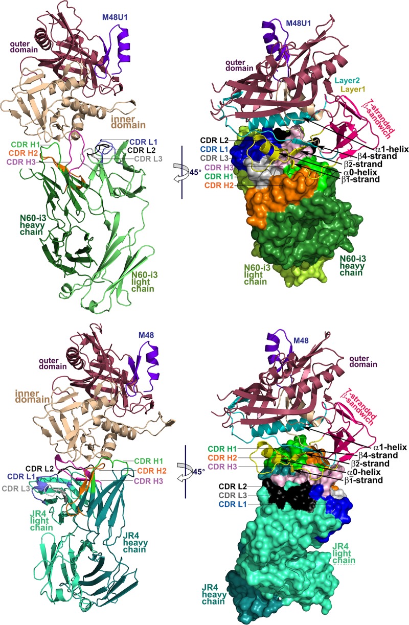
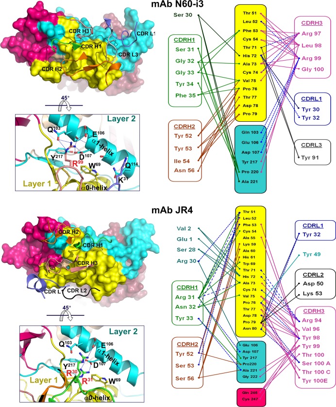
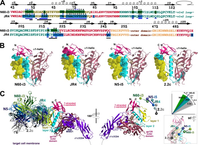
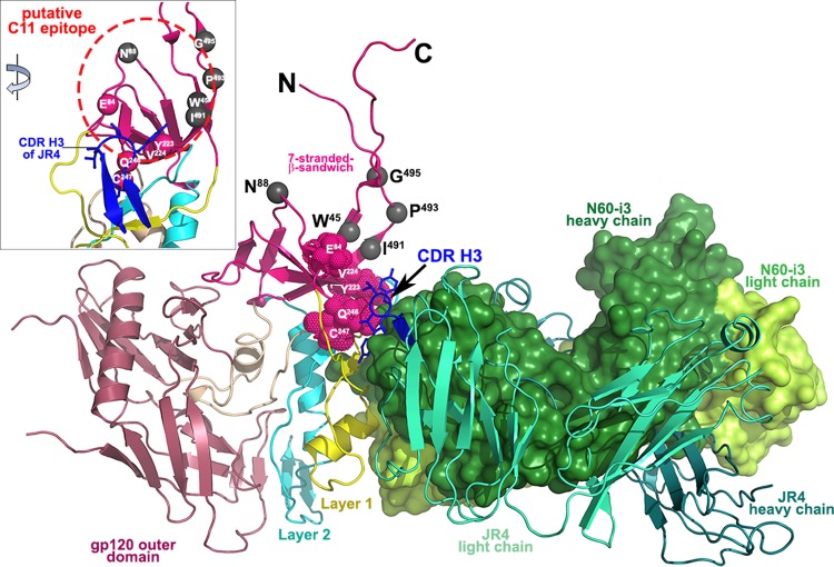
Accumulating evidence indicates a role for Fc receptor (FcR)-mediated effector functions of antibodies, including antibody-dependent cell-mediated cytotoxicity (ADCC), in prevention of human immunodeficiency virus type 1 (HIV-1) acquisition and in postinfection control of viremia. Consequently, an understanding of the molecular basis for Env epitopes that constitute effective ADCC targets is of fundamental interest for humoral anti-HIV-1 immunity and for HIV-1 vaccine design. A substantial portion of FcR effector function of potentially protective anti-HIV-1 antibodies is directed toward nonneutralizing, transitional, CD4-inducible (CD4i) epitopes associated with the gp41-reactive region of gp120 (cluster A epitopes). Our previous studies defined the A32-like epitope within the cluster A region and mapped it to the highly conserved and mobile layers 1 and 2 of the gp120 inner domain within the C1-C2 regions of gp120. Here, we elucidate additional cluster A epitope structures, including an A32-like epitope, recognized by human monoclonal antibody (MAb) N60-i3, and a hybrid A32-C11-like epitope, recognized by rhesus macaque MAb JR4. These studies define for the first time a hybrid A32-C11-like epitope and map it to elements of both the A32-like subregion and the seven-layered β-sheet of the gp41-interactive region of gp120. These studies provide additional evidence that effective antibody-dependent effector function in the cluster A region depends on precise epitope targeting--a combination of epitope footprint and mode of antibody attachment. All together these findings help further an understanding of how cluster A epitopes are targeted by humoral responses.

Importance: HIV/AIDS has claimed the lives of over 30 million people. Although antiretroviral drugs can control viral replication, no vaccine has yet been developed to prevent the spread of the disease. Studies of natural HIV-1 infection, simian immunodeficiency virus (SIV)- or simian-human immunodeficiency virus (SHIV)-infected nonhuman primates (NHPs), and HIV-1-infected humanized mouse models, passive transfer studies in infants born to HIV-infected mothers, and the RV144 clinical trial have linked FcR-mediated effector functions of anti-HIV-1 antibodies with postinfection control of viremia and/or blocking viral acquisition. With this report we provide additional definition of the molecular determinants for Env antigen engagement which lead to effective antibody-dependent effector function directed to the nonneutralizing CD4-dependent epitopes in the gp41-reactive region of gp120. These findings have important implications for the development of an effective HIV-1 vaccine.


Figures

FIG 1
FIG 1
FRET histograms of donor (A488)-labeled Fabs, acceptor (A568)-labeled Fabs, and full-length single-chain gp120BaL-CD4 complex (FLSC) in solution as determined by the FRET-FCS approach (see Materials and Methods for details). Data in panels A and B indicate ∼20% FRET efficiency for A488-labeled C11 Fab and A568-labeled A32 Fab or N60-i3 Fab bound to FLSC, respectively. Panels D and E do not show detectable FRET signals for A488-labeled A32 Fab and A568-labeled N60-i3 Fab or JR4 Fab in the presence of FLSC in solution. From the FCS measurements, the diffusion coefficient of dye-labeled Fab is ∼83 μm2/s. This diffusion has been significantly decreased upon binding two Fabs to FLSC. The diffusion coefficient for this immune complex is ∼36 μm2/s.
FIG 2
FIG 2
MAb A32/C11 competition of N60-i3/JR4 Fab binding as measured by a surface plasmon resonance competition assay. MAb A32 (A and B) and MAb C11 (B, C, and D) were immobilized on a protein A chip, and FLSC antigen was loaded to form a competitor antibody-antigen complex. Various concentrations of Fab MAb were tested. N60-i3 (A and C), JR4 (B and D), and A32 (E) were then passed over the chip. (F) Sensorgrams of the concentration series are shown. Binding affinities for MAbs A32, N60-i3, and JR4 to FLSC in the absence (blue bars) and presence (red bars) of C11 IgG were calculated with the BIAevaluation software.
FIG 3
FIG 3
Representative ADCC curves for MAbs N60-i3 and JR4. ADCC assays were performed as described in Materials and Methods using CEM-NKr-CCR5 target cells sensitized with gp120 of HIV-1BaL isolate (A) or spinoculated with AT-2-inactivated BaL viruses (B). MAb palivizumab (Synagis; MedImmune, Inc.) was included as a control.
FIG 4
FIG 4
Crystal structures of N60-i3 Fab-gp12093TH057 coree-M48U1 and JR4 Fab-gp12093TH057 coree-M48 complex. Complexes are shown in ribbon representation (right panels), or the molecular surface is displayed over Fab molecules (left panels), with the light/heavy chain of N60-i3 Fab and JR4 Fab shown in light green/dark green and light cyan/dark cyan, respectively. The complementarity-determining regions (CDRs) are shown in slate (CDR L1), black (CDR L2), gray (CDR L3), green (CDR H1), orange (CDR H2), and pink (CDR H3). The gp120 outer domain is shown in raspberry, and the inner domain is shown in wheat (left panels) or colored in a layered color scheme (right panels), with the seven-stranded β-sandwich in magenta, layer 1 in yellow, layer 2 in cyan, and layer 3 in light wheat. The mimetic peptides M48U1 and M48 are shown in violet.
FIG 5
FIG 5
Binding of MAbs N60-i3 and JR4 to gp120 antigen. On the left, the N60-i3 Fab-gp12093TH057 coree and the JR4 Fab-gp12093TH057 coree interfaces are shown with the molecular surfaces displayed at the gp120 molecules and with CDRs of Fabs shown as ribbons. Only CDRs contributing to the binding are shown. The 45° rotations show close-up views of the binding interfaces of Fabs and the α1-helix of layer 2. H bonds are shown as dashes in blue. Colors are as defined in the legend of Fig. 4. On the right, networks of interactions formed between Fabs and gp120 antigen as defined by a 5-Å distance criterion cutoff are shown as solid lines. H bonds are shown as dashes in blue.
FIG 6
FIG 6
Structural basis for interaction of cluster A MAbs with gp120 antigen. (A) Mapping of the N60-i3 and JR4 contact residues on the primary sequence of the gp120 inner domain of the 93TH057 isolate. The topology diagram depicting a distribution of secondary structure elements is shown above the gp120 sequences. Buried residues are highlighted in green and blue. Main chain (o), side chain (+), and both side and main chain (*) interactions are shown immediately above the residues as defined by a 5-Å distance criterion cutoff and colored based on contact type: hydrophobic, blue; hydrophilic, green; or both, black. Residues forming the N5-i5 and 2.2c epitopes as described in reference are indicated by blue and gray lines below the gp120 sequence, respectively. (B) Epitope footprints of MAb N60-i3, JR4, N5-i5, and 2.2c. The Cα atoms of the gp120 residues involved in Fab binding are shown as spheres and displayed over the gp120 ribbon diagram. The selected residues of layer 2 and of the seven-stranded β-sandwich contribution to Fab binding and all the residues in the α1-helix involved in N60-i3, JR4, and N5-i5 binding are labeled. (C) Comparison of binding of MAb N60-i3, JR4, N5-i5, and 2.2c to CD4-triggered gp120 antigen. The N60-i3 Fab-gp12093TH057 coree-M48, JR4 Fab-gp12093TH057 coree-M48U1, and 2.2c Fab-gp120YU2 coree-M48U1 complexes were superimposed based on the gp120 outer domain onto the N5-i5 Fab-gp12093TH057 coree-d1d2CD4 complex and oriented relative to the target cell membrane. In the 180° view only the gp12093TH057 coree and d1d2CD4 from N5-i5 Fab-gp12093TH057 coree-d1d2CD4 complex (53) are shown, with variable heavy and light (VH and VL) domains of Fabs displayed as balls. Insets show rotation angles calculated using gp120's center of mass as an origin and the average α-carbon position for the heavy chain framework region 2 (residues 36 to 49) as a reference point for each antibody (top) and a 90°rotation of the 180°view (bottom).
FIG 7
FIG 7
Putative binding site of MAb C11. The crystal structures of N60-i3 Fab-gp12093TH057 coree-M48 and JR4 Fab-gp12093TH057 coree-M48U1 are superimposed onto the HIV-1 gp120 with the gp41-interactive region (PDB code 3JWD) (72). Only the Fabs of complexes are shown, and a molecular surface is displayed over the N60-i3 Fab. The CDR H3 of JR4 predicted to block MAb C11 binding is shown in blue. The residue contacts of CDR H3 on the seven-stranded β-sandwich are shown as red spheres. Residues shown by mutagenesis studies to affect binding of MAb C11 to gp120 Env are shown as gray spheres. The inset shows a 45° rotation of the assembly where only gp120 and the CDR H3 of JR4 are shown, and the putative C11 epitope is encircled in red.

References

    1. Su B, Moog C. 2014. Which antibody functions are important for an HIV vaccine? Front Immunol 5:289. doi:10.3389/fimmu.2014.00289. - DOI - PMC - PubMed
    1. Excler JL, Ake J, Robb ML, Kim JH, Plotkin SA. 2014. Nonneutralizing functional antibodies: a new “old” paradigm for HIV vaccines. Clin Vaccine Immunol 21:1023–1036. doi:10.1128/CVI.00230-14. - DOI - PMC - PubMed
    1. Forthal D, Hope TJ, Alter G. 2013. New paradigms for functional HIV-specific nonneutralizing antibodies. Curr Opin HIV AIDS 8:393–401. doi:10.1097/COH.0b013e328363d486. - DOI - PMC - PubMed
    1. Lewis GK. 2014. Role of Fc-mediated antibody function in protective immunity against HIV-1. Immunology 142:46–57. doi:10.1111/imm.12232. - DOI - PMC - PubMed
    1. Parren PW, Marx PA, Hessell AJ, Luckay A, Harouse J, Cheng-Mayer C, Moore JP, Burton DR. 2001. Antibody protects macaques against vaginal challenge with a pathogenic R5 simian/human immunodeficiency virus at serum levels giving complete neutralization in vitro. J Virol 75:8340–8347. doi:10.1128/JVI.75.17.8340-8347.2001. - DOI - PMC - PubMed

Publication types

MeSH terms