Review
. 2015 Jul;266(1):190-207.
doi: 10.1111/imr.12309.

Linear ubiquitination in immunity

Affiliations
Review

Linear ubiquitination in immunity

Yutaka Shimizu et al. Immunol Rev. 2015 Jul.

Abstract

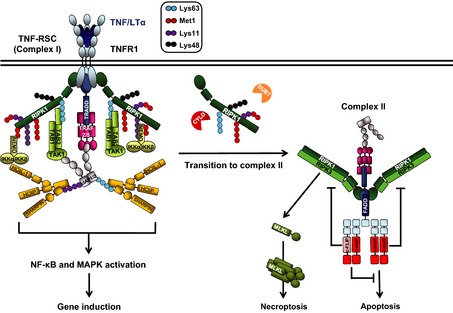
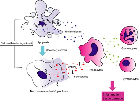
Linear ubiquitination is a post-translational protein modification recently discovered to be crucial for innate and adaptive immune signaling. The function of linear ubiquitin chains is regulated at multiple levels: generation, recognition, and removal. These chains are generated by the linear ubiquitin chain assembly complex (LUBAC), the only known ubiquitin E3 capable of forming the linear ubiquitin linkage de novo. LUBAC is not only relevant for activation of nuclear factor-κB (NF-κB) and mitogen-activated protein kinases (MAPKs) in various signaling pathways, but importantly, it also regulates cell death downstream of immune receptors capable of inducing this response. Recognition of the linear ubiquitin linkage is specifically mediated by certain ubiquitin receptors, which is crucial for translation into the intended signaling outputs. LUBAC deficiency results in attenuated gene activation and increased cell death, causing pathologic conditions in both, mice, and humans. Removal of ubiquitin chains is mediated by deubiquitinases (DUBs). Two of them, OTULIN and CYLD, are constitutively associated with LUBAC. Here, we review the current knowledge on linear ubiquitination in immune signaling pathways and the biochemical mechanisms as to how linear polyubiquitin exerts its functions distinctly from those of other ubiquitin linkage types.

Keywords: cell death; deubiquitinases; inflammation; linear ubiquitination; signaling pathways.


Figures

Figure 1
Figure 1
TNFR 1 signaling complexes and outputs. Upon TNF/LTα ligation, TNFR1 trimerizes and recruits TNFRSC (complex I) components to activate nuclear factor‐κB (NF‐κB) and MAPK signaling pathways. Different types of ubiquitination (indicated in a graphic legend) coordinate the stability of complex I. Linear ubiquitin chain assembly complex (LUBAC) is recruited to complex I to ubiquitinate RIPK1 and NF‐κB essential modulator (NEMO). Some components of complex I can dissociate and form a secondary complex, complex II. Deubiquitination of Lys63‐linked polyubiquitin on RIPK1 by cylindromatosis (CYLD) favors the transition. Other deubiquitinases (DUBs) can possibly contribute to the transition in a similar manner. Formation of complex II can result in different outcomes. First, when gene activation properly occurs, cFLIP can bind to caspase‐8 (casp8) and block cell death. Second, when gene activation is impaired, caspase‐8 forms a heterodimer and activates itself to elicit apoptosis. By contrast, when caspase‐8 activity is inhibited or when caspase‐8 or FADD is absent, RIPK1 and RIPK3 are activated and RIPK3 phosphorylates mixed lineage kinase domain‐like protein (MLKL), resulting in necroptosis.
Figure 2
Figure 2
Cell death induces inflammation. Several external stimuli can trigger different types of cell death. Necrosis and pyroptosis compromise plasma membrane integrity, which leads to the release of damage‐ (or danger‐) associated molecular patterns (DAMPs). Although apoptotic cells normally release limited DAMPs, they can produce cytokines and chemokines (so‐called find me signals), which can also attract immune cells. However, when apoptotic cells are not engulfed by phagocytes in a timely manner, which can occur when apoptosis induction is too massive or too rapid, they undergo secondary necrosis, and can hence also release DAMPs. These activate inflammatory cells which in turn secrete factors that attract and activate other types of immune cells. In summary, cell death can induce an inflammatory response which can result in tissue damage, hence autoinflammation and possibly also autoimmunity.

References

    1. Kulathu Y, Komander D. Atypical ubiquitylation – the unexplored world of polyubiquitin beyond Lys48 and Lys63 linkages. Nat Rev Mol Cell Biol 2012;13:508–523. - PubMed
    1. Glickman MH, Ciechanover A. The ubiquitin‐proteasome proteolytic pathway: destruction for the sake of construction. Physiol Rev 2002;82:373–428. - PubMed
    1. Schulman BA, Harper JW. Ubiquitin‐like protein activation by E1 enzymes: the apex for downstream signalling pathways. Nat Rev Mol Cell Biol 2009;10:319–331. - PMC - PubMed
    1. Ye Y, Rape M. Building ubiquitin chains: E2 enzymes at work. Nat Rev Mol Cell Biol 2009;10:755–764. - PMC - PubMed
    1. Li W, et al. Genome‐wide and functional annotation of human E3 ubiquitin ligases identifies MULAN, a mitochondrial E3 that regulates the organelle's dynamics and signaling. PLoS ONE 2008;3:e1487. - PMC - PubMed

Publication types