. 2015 Jun 18;522(7556):335-9.
doi: 10.1038/nature14514.

Activating positive memory engrams suppresses depression-like behaviour

Affiliations

Activating positive memory engrams suppresses depression-like behaviour

Steve Ramirez et al. Nature. .

Abstract

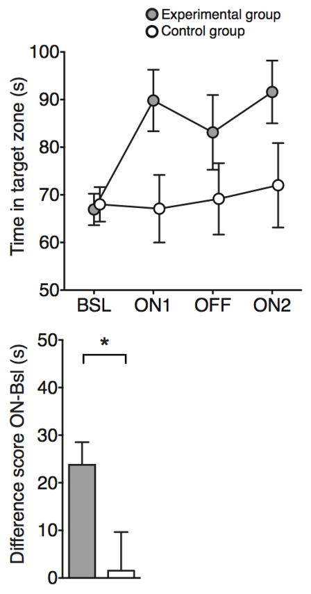
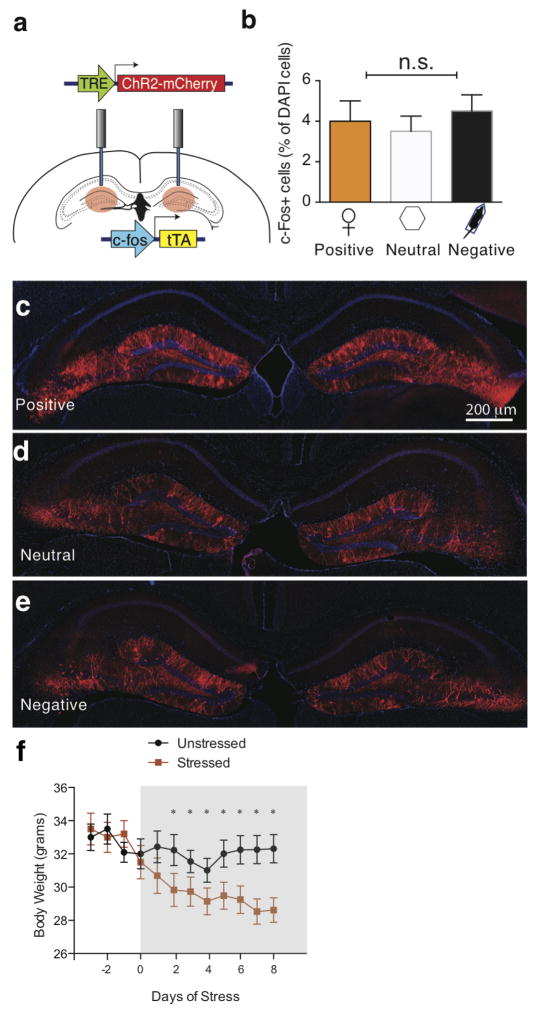
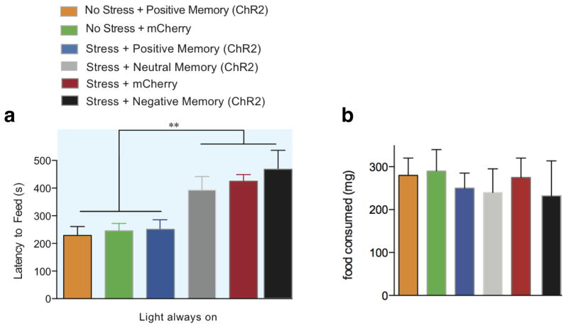
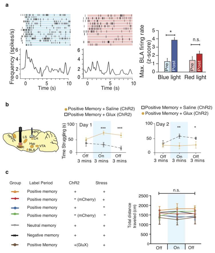
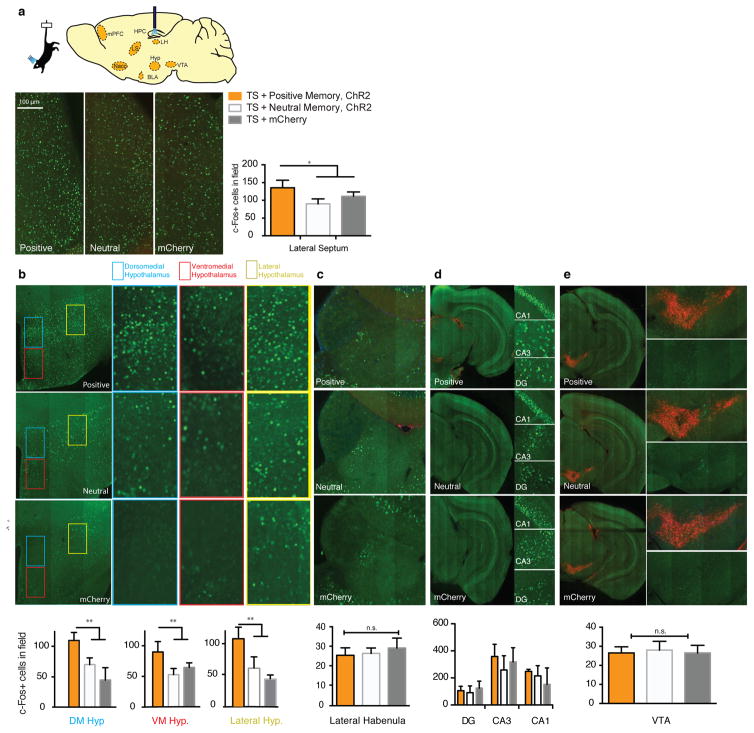
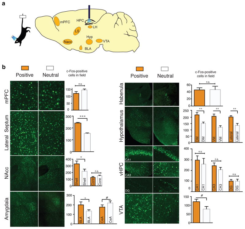
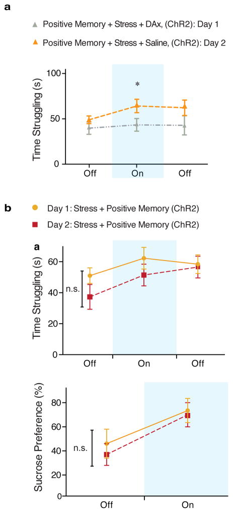
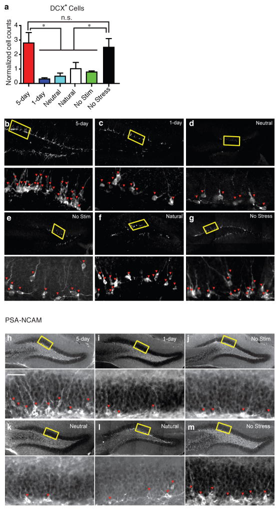
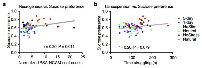
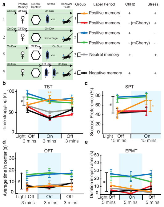
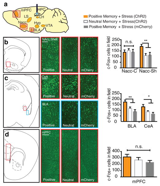
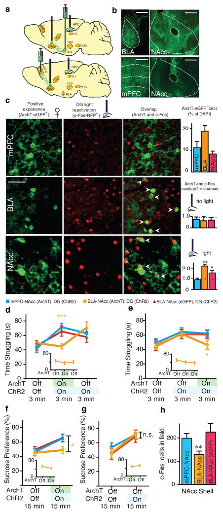
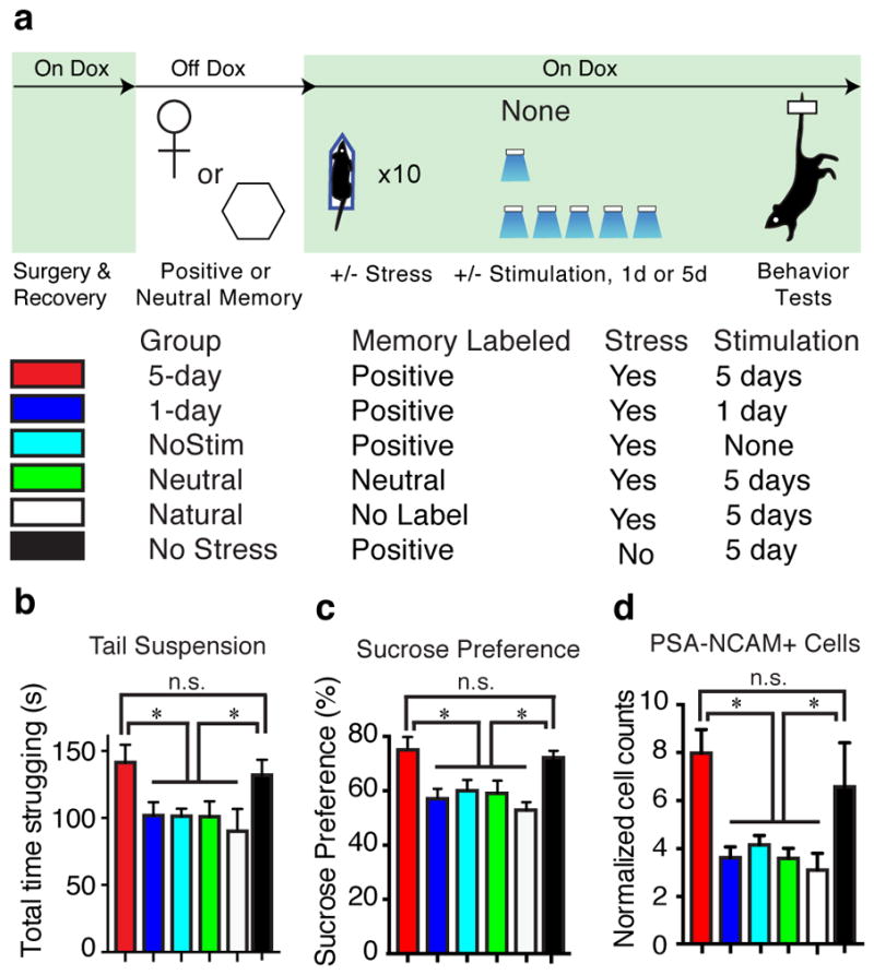
Stress is considered a potent environmental risk factor for many behavioural abnormalities, including anxiety and mood disorders. Animal models can exhibit limited but quantifiable behavioural impairments resulting from chronic stress, including deficits in motivation, abnormal responses to behavioural challenges, and anhedonia. The hippocampus is thought to negatively regulate the stress response and to mediate various cognitive and mnemonic aspects of stress-induced impairments, although the neuronal underpinnings sufficient to support behavioural improvements are largely unknown. Here we acutely rescue stress-induced depression-related behaviours in mice by optogenetically reactivating dentate gyrus cells that were previously active during a positive experience. A brain-wide histological investigation, coupled with pharmacological and projection-specific optogenetic blockade experiments, identified glutamatergic activity in the hippocampus-amygdala-nucleus-accumbens pathway as a candidate circuit supporting the acute rescue. Finally, chronically reactivating hippocampal cells associated with a positive memory resulted in the rescue of stress-induced behavioural impairments and neurogenesis at time points beyond the light stimulation. Together, our data suggest that activating positive memories artificially is sufficient to suppress depression-like behaviours and point to dentate gyrus engram cells as potential therapeutic nodes for intervening with maladaptive behavioural states.


Conflict of interest statement

The authors declare no competing financial interests.

Figures

Extended Data Figure 1
Extended Data Figure 1. Male mice spend more time around an object associated with females
a, Time spent in the target zone where the object associated with females is introduced in the ON phases. Female-object mice (experimental group) spend more time in the target zone during the ON phases than the Neutral-object mice (control group; two-way ANOVA with multiple comparisons, ON1 t88 = 2.41; p < 0.05, ON2 t88 = 2.08; p < 0.05). b, Difference score (avg of ON phases – Bsl) also shows the increased preference for the target zone in the female-object group compared to neutral-object group (t22 = 2.37; p < 0.05). n = 12 per group. See Supplementary Information for detailed methods.
Extended Data Figure 2
Extended Data Figure 2. Positive, neutral, or negative experiences label a similar proportion of dentate gyrus cells with ChR2; stress prevents weight gain over 10 days
a, The c-Fos mice were bilaterally injected with AAV9-TRE-ChR2-mCherry and implanted with optical fibers targeting DG. b–e, Histological quantifications reveal that, while off Dox, a similar proportion of DG cells are labeled by ChR2-mCherry in response to positive (c), neutral (d), or negative experience (e). All animals were sacrificed a day after completing the CIS protocol. One-way ANOVA followed by Bonferroni post-hoc test, P > 0.05, n.s. not significant. f, Animals were chronically immobilized for 10 days, during which they lost a significant amount of weight compared to an unstressed group (one-way ANOVA followed by Bonferroni post-hoc test, *P<0.05, n = 9 per group). Data are means +/− SEM.
Extended Data Figure 3
Extended Data Figure 3. Reactivation of a positive memory decreases latency to feed in a novelty suppressed feeding paradigm
a, All groups were food deprived for 24 hours and then underwent a novelty suppressed feeding protocol. While chronic immobilization increased the latency to feed, light-reactivation of a positive memory significantly decreased the latency to feed and at levels that matched the unstressed groups. b, Upon completion of the novelty suppressed feeding test, all groups were returned to their home cage and food intake was measured after 5 minutes (one-way ANOVA followed by Bonferroni post-hoc test, **P<0.01, n = 16 per group). Data are means +/− SEM.
Extended Data Figure 4
Extended Data Figure 4. Activation of a positive memory elicits BLA spiking activity, requires NAcc glutamatergic activity in the tail suspension test, but does not alter locomotor activity in the open field test
a, Raster plots and peri-stimulus time histograms (PSTH) illustrating a transient excitatory response from a single BLA neuron out of the 9 neurons responsive to DG positive memory activation during 10 seconds of blue light stimulation, but not in response to 10 seconds of red light as a control. Blue bar plots on the right illustrate maximum BLA neural firing rate before (Pre) and after (Post) blue light stimulation in the DG (paired t-test, t(7) = 6.91, P = 0.023. Red bar plots show the maximum neural activity for the same neurons after red light stimulation in the DG that serves as a control (paired t-test, t(7) = 1.62, P = 0.15). b, Brain diagram illustrating target areas manipulated. Within-subjects experiments revealed that glutamatergic antagonists (Glux), but not saline, in the accumbens shell blocked the light-induced effects of a positive memory in stressed subjects. c, All groups failed to show significant changes in locomotor activity within a session of open field exploration during either light off or light on epochs, though any trends towards decreases in locomotion are consistent with stress-induced behavioral impairments For behavioral data, a two-way ANOVA with repeated measures followed by Bonferroni post-hoc test revealed a group-by-light epoch interaction on day 1 (F1, 90 = 28.39, P < 0.001; n = 16 per group) and day 2 of testing (F1, 90 = 8.28, P < 0.01). Data are means +/− SEM.
Extended Data Figure 5
Extended Data Figure 5. Activating a positive memory in the dentate gyrus produces an increase in c-Fos expression in the lateral septum and hypothalamus, but not the lateral habenula, ventral hippocampus, or VTA
a, Diagram of regions analyzed. b, c-Fos expression significantly increased in the lateral septum (b) and subregions of the hypothalamus (c) in the positive memory group but not in a group in which a neutral memory was stimulated or in a group expressing mCherry alone. c–e, c-Fos expression did not significantly increase in the lateral habenula (c), various ventral hippocampus subregions (d), or VTA, identified by tyrosine hydroxylase staining (red) stainings in the images expanded on the right (e) (one-way ANOVA followed by Bonferroni post-hoc test *P<0.05, **P<0.01, ***P<0.001, n = 5 per group, 3–5 slices per animal). TS, tail suspension. Data are means +/− SEM.
Extended Data Figure 6
Extended Data Figure 6. Activating a positive memory through the dentate gyrus of unstressed animals increases c-Fos expression in various downstream regions
a, Diagram of regions analyzed. b, In the positive compared to the neutral memory group, c-Fos expression is significantly increased in the lateral septum, NAcc shell, BLA, dorsomedial, ventromedial, and lateral hypothalamus, but not in the mPFC, NAcc core, habenula, or ventral hippocampus. Trends were observed in the CeA and VTA. Each brain region was analyzed using an unpaired Student’s t test, n = 5 per group, 3–5 slices per animal; #P= 0.17 for CeA and P=0.09 for VTA; *P<0.05, **P<0.01, ***P<0.001, n.s. not significant. Data are means +/− SEM.
Extended Data Figure 7
Extended Data Figure 7. Dopamine receptor antagonists block the light-induced effects of positive memory activation; a single session of activating a positive memory in the dentate gyrus does not produce long-lasting antidepressant-like effects
a, Administration of a cocktail of dopamine receptor antagonists prevented the light-induced increases in struggling during the tail suspension test. When animals were tested again on day 2 and infused with saline, the behavioral effects of optically reactivating a positive memory were observed (two-way ANOVA with repeated measures followed by Bonferroni post-hoc test, *P<0.05, n = 9 per group). b, Animals in which a positive memory was optically activated during the tail suspension test or sucrose preference test showed acute increases in time struggling or preference for sucrose; this change in behavior did not persist when tested again on day 2 (within subjects ANOVA followed by Bonferroni post-hoc test), n = 9. n.s. not significant. Data are means +/− SEM.
Extended Data Figure 8
Extended Data Figure 8. Chronic activation of a positive memory prevents stress-induced decreases in neurogenesis
a, The 5-day positive memory stimulation group showed a significant increase of adult new-born cells in the DG as measured by doublecortin (DCX)-positive cells (one-way ANOVA followed by Bonferroni post-hoc test, F5, 72 = 7.634, P < 0.01) relative to control groups.. bg, Representative images of DCX-positive cells in the DG for the 5-day (b), 1-day (c), Neutral (d), No Stimulation (e), Natural (f), and No Stress (g). hm, Representative PSA-NCAM images corresponding to data appearing in Figure 4d. n = 5 slices per animal, 13 animals per group for data appearing in (a and b). *P<0.05, n.s. not significant. Data are means +/− SEM.
Extended Data Figure 9
Extended Data Figure 9. Behavioral and neuronal correlations
a, Performance levels in the SPT and the number of adult-born neurons as measured by PSA-NCAM are positively correlated on an animal-by-animal basis. b, Performance levels between the TST and SPT show strong positive correlation trends on an animal-by-animal basis. n = 14 per TST behavioral group, n = 15 per SPT behavioral group.
Figure 1
Figure 1
Activating positive memory engrams rescues depression-related behavior. a Behavior schedule and groups used. b–e, Optical reactivation of dentate gyrus (DG) cells that were previously active during a positive experience significantly increases time struggling in the tail suspension test (b) and preference for sucrose (c), but does not have a significant effect in anxiety-like behavior in the open field test (d) or elevated plus maze test (e). A two-way analysis of variance (ANOVA) with repeated measures revealed a group-by-light epoch interaction in the TST (F5, 294 = 21.20, P < 0.001) or SPT (F5, 196 = 6.20, P < 0.001) followed by Bonferroni post-hoc tests, which revealed significant increases in struggling or preference for sucrose in the positive memory + stress group. #P < 0.01. # used to denote significant differences between the four stressed groups (n = 18 per group) vs. the two non-stressed groups (n = 16 per group); *P<0.05, **P<0.01 (orange * and ** used to denote significant differences between the stress + positive memory group vs. the other three stressed groups). Data are means +/− SEM.
Figure 2
Figure 2
Positive memory reactivation increases c-Fos expression in the nucleus accumbens shell and the amygdala. a, Brain diagram illustrating target areas analyzed. b, Activation of a positive memory, but not a neutral memory or mCherry only, in the DG during the TST elicits robust c-Fos expression in the nucleus accumbens shell (b), basolateral amygdala, and central amygdala (c), but not in the medial prefrontal cortex (d). For histological data, a one-way ANOVA followed by a Bonferroni post-hoc test revealed a significant increase of c-Fos expression in the positive memory + stress group relative to controls in the NAcc and amygdala, but not the mPFC (NAcc Shell: F2, 30 = 15.2, P < 0.01; BLA: F2, 30 = 11.71, P < 0.01; CeA: F2, 30 = 11.45, P < 0.05; mPFC: F2, 30 = 1.33, P = 0.294. n = 6 per group, 3–5 slices per animal). n.s., not significant, *P<0.05, **P<0.01,***P < 0.001. Data are means +/− SEM. Scale bars correspond to 100μm.
Figure 3
Figure 3
The antidepressant effects of an optically activated positive memory require real-time terminal activity from the BLA to the NAcc. a, Brain diagram illustrating target areas manipulated. b, Representative coronal slices showing TRE-ArchT-eGFP-positive cells in the BLA or mPFC, as well as their corresponding terminals in the NAcc. Scale bars: BLA and mPFC: 500μm, NAcc, 200μm. c, Animals were taken off Dox and initially exposed to a positive experience, which caused labeling of corresponding BLA (~18%), mPFC (~12%), or NAcc (~9%) cells with eGFP derived from AAV9-TRE-ArchT-eGFP (green, halo-like expression). Light-activation of a positive memory engram in the DG preferentially reactivated the BLA and NAcc shell cells, as measured by endogenous c-Fos expression (red, nucleus-localized), that were originally labeled by the same positive experience, while no-light stimulation groups showed levels of overlap not significantly different from chance. Arrowheads indicate double-stained cells. Scale bar: 5 μm. dg, ArchT-mediated inhibition of BLA, but not mPFC, terminals in the NAcc prevents the DG-mediated light-induced increases in struggling (d, e) or preference for sucrose (f, g), while inhibition of BLA terminals in the NAcc without DG stimulation does not affect behavior (insets). h, ArchT- mediated inhibition of BLA, but not mPFC, terminals prevents the DG-mediated light-induced increase of c-Fos expression in the NAcc. For behavioral data, a two- way ANOVA with repeated measures followed by Bonferroni post-hoc test revealed a group-by-light epoch interaction and significant ArchT-mediated attenuation of struggling in the TST (d: F2, 99 = 7.30, P < 0.001; e: F2, 99 = 6.61, P < 0.01) or preference for sucrose water in the the SPT (f: F2, 66 = 10.66, P < 0.01). n = 12 per behavioral group. *P<0.05, **P<0.01, ***P<0.001; orange ** and *** used to denote significant differences between the stress + positive memory group vs. all other groups. For histological data, one-sample t-tests against chance overlap were performed (n = 4 per group, 3–5 slices per animal). n.s., not significant. Data are means +/− SEM.
Figure 4
Figure 4
Chronic activation of a positive memory elicits a long-lasting rescue of depression-related behavior. a, Behavioral schedule and groups utilized. b, c, Animals in which a positive memory was reactivated twice a day for five days showed increased struggling in a 6-minute tail suspension test (F5, 78 = 3.34, P < 0.05) (b) and increased preference for sucrose measured over 24 hours (F5, 84 = 6.25, P < 0.01) (c). d, The 5-day positive memory stimulation group showed a significant increase of adult new-born cells in the DG as measured by PSA-NCAM+ cells (F5, 72 = 4.65, P < 0.01; See Extended Data Fig. 8 for doublecortin data and PSA-NCAM images). For these data (bd), a one-way ANOVA revealed a significant interaction of the experimental-group factor and stimulation-condition factor and was followed by a Bonferroni post-hoc test. n = 14 per TST behavioral group, n = 15 per SPT behavioral group, n = 5 slices per animal for data appearing in (d). Scale bars in expanded images correspond to 5 μm. *P<0.05. Data are means +/− SEM.

Comment in

Similar articles

Cited by

References

    1. Caspi A, et al. Influence of life stress on depression: moderation by a polymorphism in the 5-HTT gene. Science. 2003;301:386–389. - PubMed
    1. Pittenger C, Duman RS. Stress, depression, and neuroplasticity: a convergence of mechanisms. Neuropsychopharmacology. 2008;33:88–109. - PubMed
    1. Hyman SE. Revitalizing psychiatric therapeutics. Neuropsychopharmacology. 2014;39:220–229. - PMC - PubMed
    1. Covington HE, III, et al. Antidepressant effect of optogenetic stimulation of the medial prefrontal cortex. The Journal of neuroscience. 2010;30:16082–16090. - PMC - PubMed
    1. Russo SJ, Nestler EJ. The brain reward circuitry in mood disorders. Nat Rev Neurosci. 2013;14:609–625. - PMC - PubMed

Publication types

MeSH terms

Substances