Review
. 2015 Jul;28(3):721-41.
doi: 10.1128/CMR.00127-14.

Diversity and Evolution in the Genome of Clostridium difficile

Affiliations
Review

Diversity and Evolution in the Genome of Clostridium difficile

Daniel R Knight et al. Clin Microbiol Rev. 2015 Jul.

Abstract

Clostridium difficile infection (CDI) is the leading cause of antimicrobial and health care-associated diarrhea in humans, presenting a significant burden to global health care systems. In the last 2 decades, PCR- and sequence-based techniques, particularly whole-genome sequencing (WGS), have significantly furthered our knowledge of the genetic diversity, evolution, epidemiology, and pathogenicity of this once enigmatic pathogen. C. difficile is taxonomically distinct from many other well-known clostridia, with a diverse population structure comprising hundreds of strain types spread across at least 6 phylogenetic clades. The C. difficile species is defined by a large diverse pangenome with extreme levels of evolutionary plasticity that has been shaped over long time periods by gene flux and recombination, often between divergent lineages. These evolutionary events are in response to environmental and anthropogenic activities and have led to the rapid emergence and worldwide dissemination of virulent clonal lineages. Moreover, genome analysis of large clinically relevant data sets has improved our understanding of CDI outbreaks, transmission, and recurrence. The epidemiology of CDI has changed dramatically over the last 15 years, and CDI may have a foodborne or zoonotic etiology. The WGS era promises to continue to redefine our view of this significant pathogen.


Figures

FIG 1
FIG 1
Circular illustration of the 4.3-Mb chromosome of C. difficile strain 630. The concentric circles are as follows (from the outside in): circles 1 and 2, 3,776 putative CDSs (transcribed clockwise and counterclockwise); circle 3, CDSs shared with other sequenced Clostridia (blue); circle 4, CDSs unique to C. difficile (red); circle 5, mobile elements (red/pale red, transposons; pink, prophages; brown, partial prophages/transposons; blue, skin element; magenta, genomic island); circle 6, RNA genes (blue, rRNAs; red, tRNAs; purple, stable RNAs); circles 7 and 8, G+C content/GC deviation (plotted using a 10-kb window). (Reproduced from reference by permission from Macmillan Publishers Ltd.)
FIG 2
FIG 2
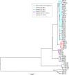
Phylogenetic tree showing representatives of six currently described C. difficile clades and the relationship between toxigenic and nontoxigenic isolates. A maximum likelihood tree was generated from the alignment of 1,426 core genes of 73 C. difficile isolates. Isolates represented extremes of clinical severity, geographic diversity, and toxigenic status. Clades are indicated by their designated number. Nontoxigenic isolates are indicated by black branches. Toxigenic isolates are indicated by branches colored according to clade. The ST and RT (in parentheses) of a well-characterized representative of each clade are indicated. (Reproduced from reference by permission of the Society for Molecular Biology and Evolution.)
FIG 3
FIG 3
Transcontinental dissemination of epidemic RT027. Shown is the global spread (arrows) of lineages FQR1 and FQR2 inferred from phylogeographic analysis. The width of the arrow is approximately proportional to the number of descendants from each sublineage. The inset shows an enlarged view of transmission in Europe. (Reproduced from reference by permission from Macmillan Publishers Ltd.)
FIG 4
FIG 4
Diverse sources of C. difficile in the hospital environment. Shown are the genetic variation and epidemiological relationships among 957 isolates obtained from patients with CDI. (A) Numbers of single-nucleotide variants (SNVs) between each sample obtained during the period from 1 April 2008 through 31 March 2011 and the most closely related previous sample obtained after 1 September 2007. (B) Percentages of isolates that were classified as genetically related, according to the different SNV thresholds, along with the epidemiological links between related isolates. (Reproduced from reference with permission from the Massachusetts Medical Society.)
None
None
None
None
None

References

    1. Rupnik M, Wilcox MH, Gerding DN. 2009. Clostridium difficile infection: new developments in epidemiology and pathogenesis. Nat Rev Microbiol 7:526–536. doi:10.1038/nrmicro2164. - DOI - PubMed
    1. Wiegand PN, Nathwani D, Wilcox MH, Stephens J, Shelbaya A, Haider S. 2012. Clinical and economic burden of Clostridium difficile infection in Europe: a systematic review of healthcare-facility-acquired infection. J Hosp Infect 81:1–14. doi:10.1016/j.jhin.2012.02.004. - DOI - PubMed
    1. Centers for Disease Control and Prevention. 2013. Antibiotic resistance threats in the United States, 2013. Centers for Disease Control and Prevention, Atlanta, GA: http://www.cdc.gov/drugresistance/threat-report-2013/.
    1. Lessa FC, Mu Y, Bamberg WM, Beldavs ZG, Dumyati GK, Dunn JR, Farley MM, Holzbauer SM, Meek JI, Phipps EC, Wilson LE, Winston LG, Cohen JA, Limbago BM, Fridkin SK, Gerding DN, McDonald LC. 2015. Burden of Clostridium difficile infection in the United States. N Engl J Med 372:825–834. doi:10.1056/NEJMoa1408913. - DOI - PMC - PubMed
    1. Hall IC, O'Toole E. 1935. Intestinal flora in newborn infants with a description of a new pathogenic anaerobe, Bacillus difficilis. Am J Dis Child 49:390–402. doi:10.1001/archpedi.1935.01970020105010. - DOI

Publication types

MeSH terms

LinkOut - more resources