Review
. 2015 Jul;28(3):801-70.
doi: 10.1128/CMR.00109-14.

Blood Groups in Infection and Host Susceptibility

Affiliations
Review

Blood Groups in Infection and Host Susceptibility

Laura Cooling. Clin Microbiol Rev. 2015 Jul.

Abstract

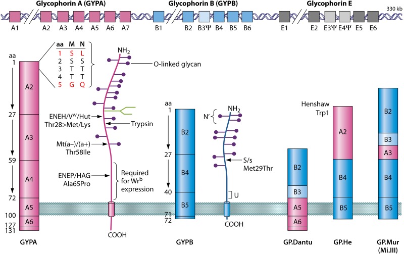
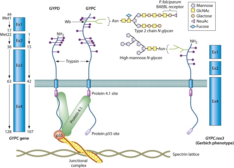
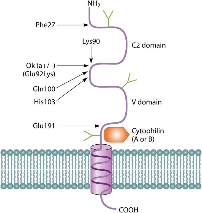
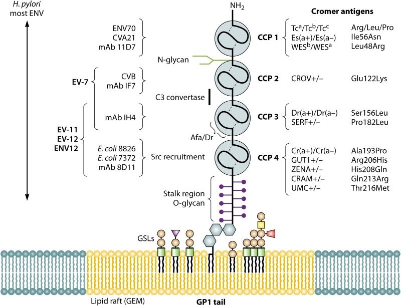
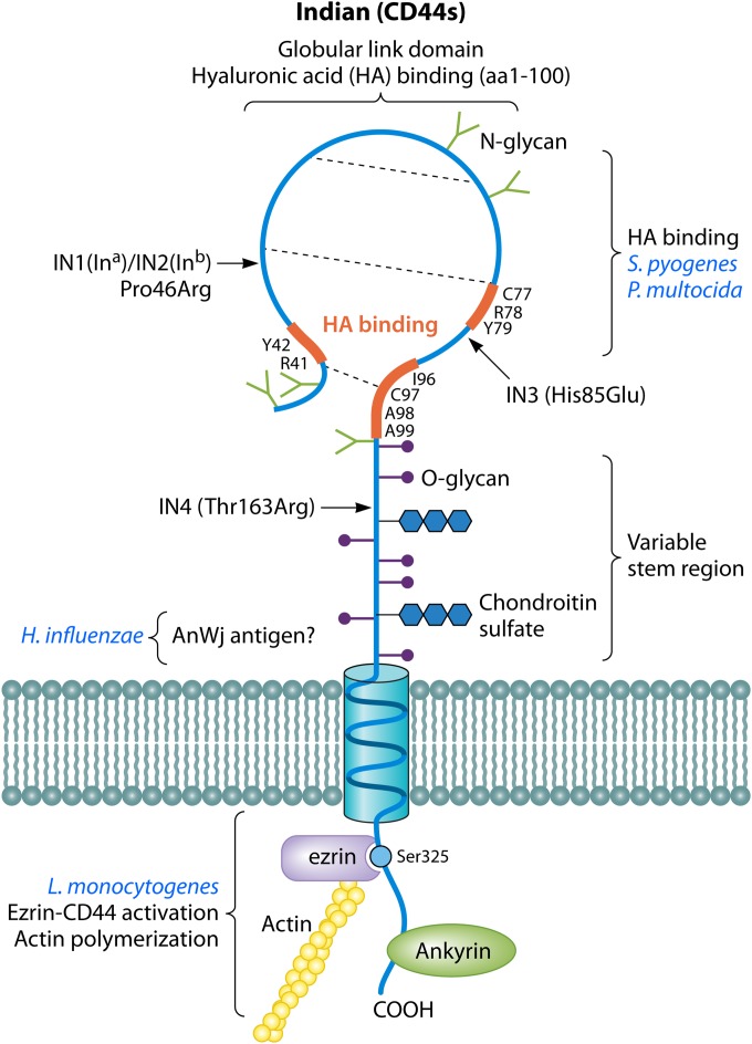
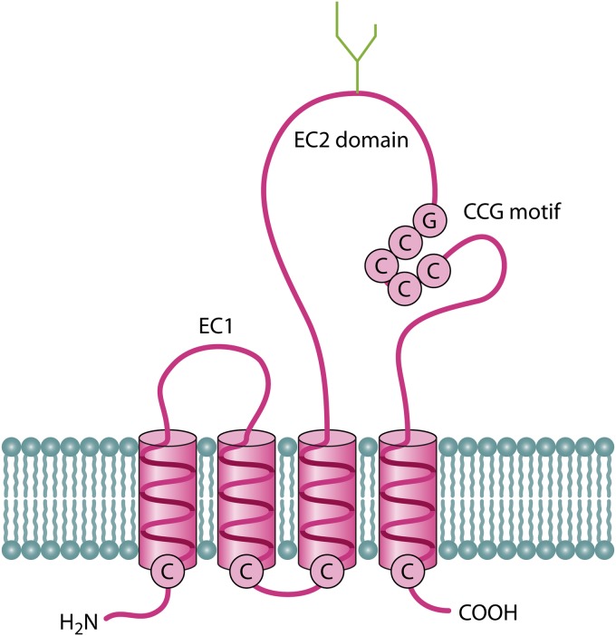
Blood group antigens represent polymorphic traits inherited among individuals and populations. At present, there are 34 recognized human blood groups and hundreds of individual blood group antigens and alleles. Differences in blood group antigen expression can increase or decrease host susceptibility to many infections. Blood groups can play a direct role in infection by serving as receptors and/or coreceptors for microorganisms, parasites, and viruses. In addition, many blood group antigens facilitate intracellular uptake, signal transduction, or adhesion through the organization of membrane microdomains. Several blood groups can modify the innate immune response to infection. Several distinct phenotypes associated with increased host resistance to malaria are overrepresented in populations living in areas where malaria is endemic, as a result of evolutionary pressures. Microorganisms can also stimulate antibodies against blood group antigens, including ABO, T, and Kell. Finally, there is a symbiotic relationship between blood group expression and maturation of the gastrointestinal microbiome.


Figures

FIG 1
FIG 1
Synthesis of H, A, and B antigens. The H antigen is formed by the addition of an α1-2 fucose (blue diamonds) by FUT1 or H-glycosyltransferase. H antigen can then serve as a substrate for ABO glycosyltransferase. Group A individuals express an α1-3 N-acetylgalactosamine (GalNAc) (red trapezoid), and group B individuals express an α1-3 galactose (Gal) (orange circles). Group O individuals have inactive ABO genes and express only the H-antigen precursor. The Bombay phenotype (Oh) lacks H, A, and B antigens due to null FUT1 alleles.
FIG 2
FIG 2
Relationship and synthesis of type 1 (Lea and Leb) and type 2 (LeX and LeY) antigens by Lewis (FUT3) and Secretor (FUT2) enzymes and their modification by ABO.
FIG 3
FIG 3
ABO gene and major A, B, and O alleles. The ABO gene resides on chromosome 9q34.1 and contains 7 exons, which encode a 354-aa glycoprotein. The glycoproteins encoded by the A (ABO*A1) and B (ABO*B) consensus alleles differ by 4 aa, 3 of which are functionally important (aa 235, 266, and 268). Group O alleles belonging to the O1 family contain a deletion in exon 6, leading to a frameshift and a premature stop codon. Alleles belonging to the O2 family share a mutation at aa 268. Shown in red are the SNP designations commonly used in genomic studies for ABO typing.
FIG 4
FIG 4
Cholera lectin binding sites. Ganglioside GM1 is the primary CTx receptor. The binding site involves residues on adjacent B subunits. A second, weaker binding site for LeY-type epitopes is located along the other face of the B subunit (yellow diamonds).
FIG 5
FIG 5
Synthesis of globo- and related neolacto- and gala-series GSLs. Also shown are agents capable of inhibiting GSL synthesis (PPMP), inducing enzyme expression (TNF and H. pylori), and inactivating mutations (Pk and p phenotypes). Microbial lectins recognizing GSL antigens are included.
FIG 6
FIG 6
Globoside and parvovirus B19. (A) Distribution of Gb4 in human tissues (mean percent total neutral GSLs ± standard deviation). HUVEC, human umbilical vein endothelial cell. (B) The parvovirus B19 capsid is composed of VP2 (light blue) and VP1 (dark blue) protomers. B19 binds to Gb4 on cell membranes (for example, red cells), inducing a conformational change in VP1 with surface expression of the VP1u peptide. The majority of the modified B19 dissociates, where it may adhere to a high-affinity coreceptor on erythroblast membranes. It is likely that the receptor lies within a Gb4-enriched microdomain that facilitates B19 uptake.
FIG 7
FIG 7
Duffy or DARC antigen and gene. (A) The DARC glycoprotein contains a large extracellular domain and 7 transmembrane domains. The amino-terminal extracellular domain contains the P. vivax PvDBP binding site (aa 8 to 42), of which the negatively charged Fy6 epitope (aa 19 to 26) is critical. The Fya/b polymorphism resides at amino acid 42. A missense mutation in the first cytoplasmic loop of the transmembrane domain (Arg89Cys) is responsible for the FyX phenotype, which is associated with weak DARC expression. (B) The DARC gene resides on chromosome 1q23.2. DARC expression on red cells is silenced in the FY*BES allele, which contains a mutation in a GATA promoter site. The SNP designations and the location of FY*BES, FY*B/FY*A, and FY*X are shown.
FIG 8
FIG 8
Diego blood group (AE1; band 3; SLC4A1). AE1 exists as dimers and tetramers and is preferentially located at junctional complexes and the “band 3-ankyrin” metabolon (shown). The amino-terminal cytoplasmic domain is involved in oligomerization and interacts with the underlying cytoskeleton. Several glycolytic enzymes and hemoglobin derivatives or hemichromes (methemoglobin and denatured hemoglobin) associate with the amino-terminal domain. Carbonic anhydrase is associated with the cytoplasmic carboxy tail. The large transmembrane domain is an anion transporter, exchanging Cl/HCO3 anions. Several high/low-incidence antigens are located along the extracellular loops. The high-incidence Wrb antigen requires interaction with glycophorin A for expression. A 27-bp deletion at the junction of the amino-terminal and transmembrane domain is responsible for Southeast Asian ovalocytosis (SLC4A1Δ27) (in red). AE1 contains a single massive N-glycan that expresses ABO and is responsible for 50% of all the ABO antigens on red blood cells.
FIG 9
FIG 9
MNSs blood group (GYPA and GYPB). Glycophorins A, B, and E are located together on chromosome 4q28-q31 and can undergo recombination, leading to hybrid glycophorins (for example, Henshaw, GP.Mur, and GP.Dantu) and null phenotypes [En(a−), U, and Mk]. The MN antigens are defined by the first 5 amino acids and O-linked glycans on glycophorin A. Glycophorin A contains 15 O-glycan sites and >20 high/low antigens. Glycophorin A is physically associated with AE1/band 3 and participates in Wrb antigen expression located on AE1. The location of 3 high/low polymorphisms showing evidence of selection (16) and the trypsin cleavage site are shown. The S/s antigens are a single polymorphism on glycophorin B. The U antigen is located along a short peptide segment near the membrane.
FIG 10
FIG 10
Gerbich blood group (GYPC and GYPD). Glycophorins C and D are products of the GYPC gene and differ by 21 amino acids at the amino-terminal end. Both proteins interact with the cytoskeleton elements at junctional complexes. GYPC contains a single N-glycan that binds the P. falciparum protein EBA140/BAEBL. The Gerbich phenotype is a deletion mutant lacking exon 3. The altered protein is underglycosylated and expresses an immature, high-mannose N-glycan. In addition, there is an altered interaction with the junctional complexes, leading to ovalocytosis.
FIG 11
FIG 11
Knops blood group (CR1; CD35). The CR1 variant on red cells is composed of 30 complement consensus repeats arranged as 4 long homologous repeats (LHR-A to -D). CCPs involved in complement binding are highlighted (purple) and referred to as sites 1, 2, and 2′. These sites also serve as binding sites for malarial proteins (PfRh4 and PfEMP). LHR-D may interact with mannose binding lectin. The Knops antigens are located in CCP25 and CCP26 (blue). The sites of two mutations associated with weak CR1/Knops expression are highlighted in green. Also shown is the trypsin cleavage site on CR1.
FIG 12
FIG 12
OK blood group (Basigin; CD147). CD147 is a member of the immunoglobulin superfamily. The most common isoform contains single C-type and V-type domains. The Ok(a+/a−) polymorphism is located at amino acid 92. Several amino acids that are believed to participate in PfRh5 binding are highlighted (612, 615). CD147 is also able to bind cyclophilins (A and B) and plays a role in many viral infections (HIV, measles virus, and SARS-CoV).
FIG 13
FIG 13
Cromer blood group. Decay-accelerating factor (DAF) (CD55) is a GPI-linked glycoprotein composed of 4 complement consensus domains (CCPs), a heavily glycosylated stalk region, and a GPI tail. The location of Cromer antigens is shown on the right. C3 convertase binding is located along CCP2-CCP3. CCP domains involved in binding specific echoviruses (EV), enteroviruses (ENV), bacteria, and MAbs are shown. H. pylori and many enteroviruses appear to require the entire molecule for binding. Like most GPI-linked glycoproteins, DAF is a raft protein and is localized at or recruited into glycolipid-enriched microdomains (GEMs).
FIG 14
FIG 14
CD44/Indian blood group. CD44s is a 341-aa glycoprotein that possesses a globular link domain with three disulfide bonds (hatched lines) and 5 to 6 N-glycans. The globular domain is capable of binding hyaluronic acid (HA), including hyaluronic acid on S. pyogenes. Peptide regions critical for HA binding are highlighted in red. The glycosylated stem region expresses both O-linked glycans (purple) and chondroitin sulfate (blue hexagons) and can vary between tissues due to alternate splicing and glycosylation. The AnWj epitope is hypothesized to lie in the stalk region and is a receptor for H. influenzae. The molecule contains a long cytoplasmic domain that interacts with ankyrin and ERM proteins to modulate the cytoskeleton. Binding by L. monocytogenes invokes ezrin binding and phosphorylation with actin polymerization.
FIG 15
FIG 15
Raph blood group (CD151; MER2). Raph is located on the tetraspanin CD151. Like all tetraspanins, CD151 is a multipass protein with two extracellular loops. The EC2 domain is functionally critical for protein function. Tetraspanins can act as receptors for viruses or may help organize receptors within tetraspanin-enriched membrane domains.
None

References

    1. Fung MK, Grossman BJ, Hillyer CD, Westhoff CM. 2014. Technical manual, 18th ed AABB Press, Bethesda, MD.
    1. Reid ME, Lomas-Francis C, Olsson ML. 2012. Blood group antigen FactsBook, 3rd ed Academic Press, Waltham, MA.
    1. Burton NM, Bruce LJ. 2011. Modeling the structure of the red cell membrane. Biochem Cell Biol 89:200–215. doi: 10.1139/O10-154. - DOI - PubMed
    1. An X, Mohandas N. 2008. Disorders of red cell membrane. Br J Haematol 141:367–375. doi: 10.1111/j.1365-2141.2008.07091.x. - DOI - PubMed
    1. Hosseini SM, Feng JJ. 2012. How malaria parasites reduce the deformability of infected RBC. Biophys J 103:1–10. doi: 10.1016/j.bpj.2012.05.026. - DOI - PMC - PubMed

MeSH terms

Substances

LinkOut - more resources