

Review
doi: 10.1523/JNEUROSCI.0409-15.2015.
Monosynaptic Circuit Tracing with Glycoprotein-Deleted Rabies Viruses
Affiliations
- PMID: 26085623
- PMCID: PMC4469731
- DOI: 10.1523/JNEUROSCI.0409-15.2015
Item in Clipboard
Review
Monosynaptic Circuit Tracing with Glycoprotein-Deleted Rabies Viruses
J Neurosci.
.
No abstract available
Figures
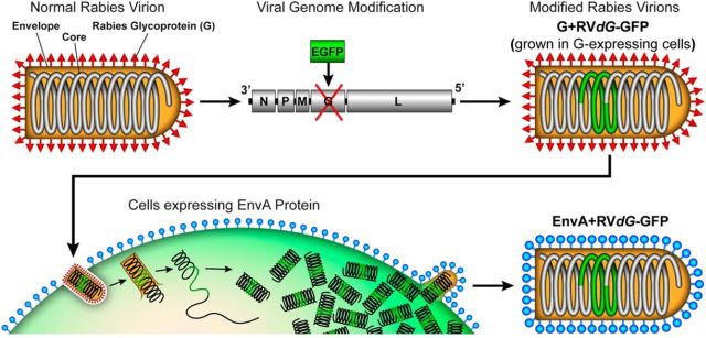
Glycoprotein deletion and EnvA pseudotyping of rabies virus. Normal rabies viral particles (top, left) are composed of a viral core surrounded by a host cell-derived membrane envelope in which the rabies glycoprotein (G; red spikes) is embedded. The core of the virus includes structural proteins and the negative strand RNA genome. The genome codes for five different proteins, including G (top, middle). The viral genome can be modified such that the coding sequence for G is deleted and can be replaced with the coding sequence for a transgene, such as GFP, which will be expressed in infected cells. G-deleted rabies virus particles (RVdG) are initially produced de novo from DNA and then can be propagated indefinitely (Osakada and Callaway, 2013). When propagated in cells that express G (data not shown), RVdG particles will bud out from the host cells with G on their envelope (G + RVdG, top, right). To pseudotype RVdG, cells expressing EnvA (blue lollipops) are infected with G + RVdG (bottom, left) and the particles that emerge will have EnvA on their envelope (bottom, right). These EnvA-pseudotyped particles are designated EnvA + RVdG.
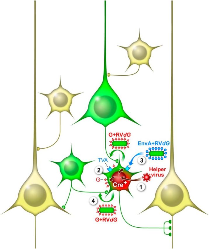
Monosynaptic rabies tracing of inputs to Cre-expressing starter cells. Monosynaptic tracing with G-deleted rabies (RVdG) involves expression of the EnvA receptor, TVA, and rabies glycoprotein (G) in starter cells. These cells are subsequently infected specifically with EnvA + RVdG, which is trans-complemented by G to produce G + RVdG that spreads trans-synaptically to neurons providing synaptic input to starter cells. The spread of RVdG is monosynaptically restricted because the input cells lack G and G is required for trans-synaptic spread. In this example, the starter cells are selected on the basis of Cre expression in a mouse line expressing Cre in a cell type of interest. In the first step (1) a helper virus that expresses TVA, G, and a marker gene (RFP) in a Cre-dependent manner is injected in the location of desired starter cells. This results in expression of TVA, G, and RFP only in the Cre+ cells (green and red) (2). After a sufficient time for accumulation of TVA and G, EnvA + RVdG is injected in the location (3) and selectively infects the TVA+ starter cells. In this example the EnvA + RVdG contains the coding sequence for GFP resulting in GFP expression in the starter cells. The RVdG replicates in the starter cells and trans-complementation with G results in the production of G + RVdG that spreads trans-synaptically to input cells (4), which express GFP from the RV genome (green). The result is that starter cells are marked with both RFP and GFP while input cells express only GFP. Other neurons that do not express Cre and are not directly presynaptic to starter cells remain unlabeled (light yellow).
References
Publication types
MeSH terms
Substances
Grants and funding
LinkOut - more resources
Full Text Sources
Other Literature Sources