. 2015 Jun 17;35(24):9078-87.
doi: 10.1523/JNEUROSCI.5043-14.2015.

Developmental Changes in Brain Network Hub Connectivity in Late Adolescence

Affiliations

Developmental Changes in Brain Network Hub Connectivity in Late Adolescence

Simon T E Baker et al. J Neurosci. .

Abstract

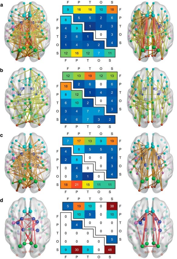
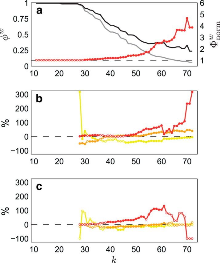
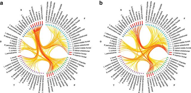
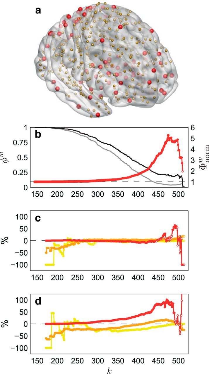
The human brain undergoes substantial development throughout adolescence and into early adulthood. This maturational process is thought to include the refinement of connectivity between putative connectivity hub regions of the brain, which collectively form a dense core that enhances the functional integration of anatomically distributed, and functionally specialized, neural systems. Here, we used longitudinal diffusion magnetic resonance imaging to characterize changes in connectivity between 80 cortical and subcortical anatomical regions over a 2 year period in 31 adolescents between the ages of 15 and 19 years. Connectome-wide analysis indicated that only a small subset of connections showed evidence of statistically significant developmental change over the study period, with 8% and 6% of connections demonstrating decreased and increased structural connectivity, respectively. Nonetheless, these connections linked 93% and 90% of the 80 regions, respectively, pointing to a selective, yet anatomically distributed pattern of developmental changes that involves most of the brain. Hub regions showed a distinct tendency to be highly connected to each other, indicating robust "rich-club" organization. Moreover, connectivity between hubs was disproportionately influenced by development, such that connectivity between subcortical hubs decreased over time, whereas frontal-subcortical and frontal-parietal hub-hub connectivity increased over time. These findings suggest that late adolescence is characterized by selective, yet significant remodeling of hub-hub connectivity, with the topological organization of hubs shifting emphasis from subcortical hubs in favor of an increasingly prominent role for frontal hub regions.

Keywords: MRI; adolescence; connectome; development; graph theory; structural connectivity.


Figures

Figure 1.
Figure 1.
Developmental changes in structural connectivity. a illustrates the anatomical arrangement of the subset of edges identified by the NBS (Zalesky et al., 2010a) as showing significant decreases (left) and increases (right) in streamline density over time, with the thickness of each connection weighted by its associated one-tailed t test statistic (FWE corrected, p < 0.05). Edge color represents connection type: peripheral (yellow), feeder (orange), and hub–hub (red), with larger nodes corresponding to hub regions. Node color represents the assignment of each region of interest to one of five broad anatomical divisions: frontal (cyan), parietal (lime), temporal (magenta), occipital (orange-red), or subcortical (blue). The center panel illustrates the anatomical distribution of the developmental decreases (lower triangular matrix) and increases (upper triangular matrix) in connectivity based on the classification of edges according to the anatomical divisions they interconnected. The values in these matrices represent relative proportions, calculated as the ratio between (1) the frequency of edges linking each pair of divisions and (2) the total number of edges belonging to the two categories. Illustrated in b–d are peripheral, feeder, and hub–hub connections, respectively, extracted from the NBS subnetworks in a.
Figure 2.
Figure 2.
Rich-club curves and magnitude of developmental changes in peripheral, feeder, and hub–hub connections. a shows the normalized rich-club coefficients, Φnormw(k) (red), for the follow-up group-averaged streamline density-weighted network. Weighted rich-club coefficients, φw(k) (black), were found to be significantly greater than those derived from randomized networks, φrandw(k) (gray), for a range of k from 30 to 72 (one-tailed, p < 0.05; filled/solid red markers), indicating robust rich-club organization in the network. b and c show the percentage difference between observed and expected proportions at each level of k for each connection type [peripheral (yellow), feeder (orange), and hub–hub (red)] in the subnetwork showing decreased streamline density over time (b) and the subnetwork showing increased streamline density over time (c). χ2 analysis of the difference between the observed and expected proportions (also at each level of k) revealed a significant effect of connection type (p < 0.05; filled/solid markers) in both the subnetwork showing decreased streamline density over time (for each k in the range 27 < k < 36 except k = 31, and 45 < k < 73; b) and the subnetwork showing increased streamline density over time (for each k in the range 30 < k < 62 except k = 54; c). As can be seen, a disproportionate number of hub–hub connections showed developmental changes, particularly at levels of k that coincide with the topological rich-club effect (a).
Figure 3.
Figure 3.
Connectograms of developmental decreases and increases in streamline density are shown in a and b, respectively, with links colored by connection type: peripheral (yellow), feeder (orange), and hub–hub (red). Regions of interest are grouped according to broad anatomical divisions [i.e., frontal (cyan), parietal (lime), temporal (magenta), occipital (orange-red), or subcortical (blue)] and are further ordered by degree (bar height is proportional to degree), with bars colored in red corresponding to hub regions. F, Frontal; L, left; O, occipital; P, parietal; R, right; S, subcortical; T, temporal.
Figure 4.
Figure 4.
The effect of parcellation scale on developmental changes in structural connectivity. a illustrates the anatomical location of nodes classified as hubs (red) and nonhubs (gold) in the follow-up group-averaged streamline density-weighted network based on the 530-node parcellation. b shows the normalized rich-club coefficients, Φnormw(k) (red), for this network. Weighted rich-club coefficients, φw(k) (black), were found to be significantly greater than those derived from randomized networks, φrandw(k) (gray), for each k in the range 143 < k < 170 and 172 < k < 510 (one-tailed, p < 0.05; filled/solid red markers), indicating robust rich-club organization in the network. c and d show the percentage difference between observed and expected proportions at each level of k for each connection type [peripheral (yellow), feeder (orange), and hub–hub (red)] in the subnetwork showing decreased streamline density over time (c) and the subnetwork showing increased streamline density over time (d). χ2 analysis of the difference between the observed and expected proportions (also at each level of k) revealed a significant effect of connection type (p < 0.05; filled/solid markers) in both the subnetwork showing decreased streamline density over time (for the majority of k in the range 171 < k < 503; c) and the subnetwork showing increased streamline density over time (for each k in the range 171 < k < 491 and 508 < k < 515; d). As can be seen, developmental changes—particularly increases—in streamline density were predominantly related to hub–hub connectivity, particularly at levels of k that coincide with the topological rich-club effect (b).

References

    1. Ahuja RK, Magnanti TL, Orlin JB. Network flows: theory, algorithms, and applications. Upper Saddle River, NJ: Prentice Hall; 1993.
    1. Ball G, Aljabar P, Zebari S, Tusor N, Arichi T, Merchant N, Robinson EC, Ogundipe E, Rueckert D, Edwards AD, Counsell SJ. Rich-club organization of the newborn human brain. Proc Natl Acad Sci U S A. 2014;111:7456–7461. doi: 10.1073/pnas.1324118111. - DOI - PMC - PubMed
    1. Benes FM, Turtle M, Khan Y, Farol P. Myelination of a key relay zone in the hippocampal formation occurs in the human brain during childhood, adolescence, and adulthood. Arch Gen Psychiatry. 1994;51:477–484. doi: 10.1001/archpsyc.1994.03950060041004. - DOI - PubMed
    1. Bullmore E, Sporns O. Complex brain networks: graph theoretical analysis of structural and functional systems. Nat Rev Neurosci. 2009;10:186–198. doi: 10.1038/nrn2575. - DOI - PubMed
    1. Colizza V, Flammini A, Serrano MA, Vespignani A. Detecting rich-club ordering in complex networks. Nat Phys. 2006;2:110–115. doi: 10.1038/nphys209. - DOI

Publication types

LinkOut - more resources