Case Reports
doi: 10.4103/0974-2069.154154.
Isolation of the right subclavian artery in a patient with d-transposition of the great arteries
Affiliations
- PMID: 26085773
- PMCID: PMC4453190
- DOI: 10.4103/0974-2069.154154
Item in Clipboard
Case Reports
Isolation of the right subclavian artery in a patient with d-transposition of the great arteries
Ann Pediatr Cardiol.
2015 May-Aug.
Abstract
Isolation of the right subclavian artery (RSCA) is rare, and this finding in association with d-transposition of the great arteries (d-TGA) is extremely unusual. We present a case of an isolated RSCA in a newborn with d-TGA in whom the clinical presentation was diagnostic. We discuss the imaging modalities used to confirm the diagnosis, the embryological basis of the finding, and the surgical repair.
Keywords: Anomalous origin of right subclavian artery; congenital heart disease; isolated right subclavian artery; transposition of the great arteries.
Conflict of interest statement
Figures
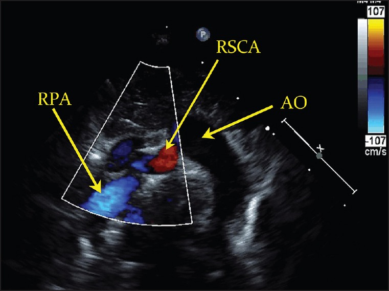
Transthoracic echocardiogram demonstrating a vessel arising from the proximal right pulmonary artery (RPA) and coursing superior and rightward, suggestive of an anomalous origin of the right subclavian artery (RSCA). AO = Aorta
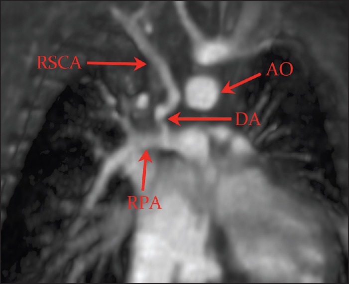
Thin-slab three-dimensional surface rendering magnetic resonance imaging confirming the isolated RSCA from a right ductus arteriosus (DA) arising from the proximal RPA
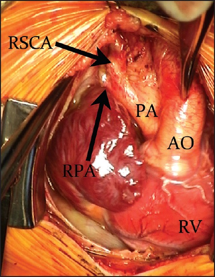
Intraoperative image of the RSCA connected to the RPA via a long segment of ductal tissue. RV = Right ventricle
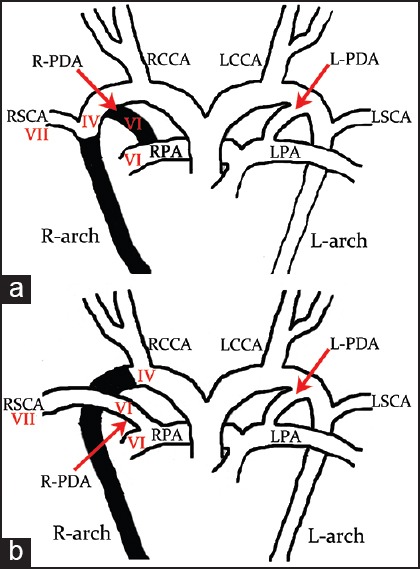
(a) Diagram of the normal embryological origin of the RSCA from the seventh (VII) intersegmental artery with subsequent cranial migration, and dissolution (in black) of the R-PDA and right fourth (IV) aortic arch (R-arch). (b) Isolation of the RSCA occurs when there is dissolution (in black) of the right IV aortic arch but persistence of the right sixth (VI) arch from which the R-PDA and RPA take their origin. L-arch = Left aortic arch, LCCA = left common carotid artery, L-PDA = left ductus arteriosus, LSVC = left subclavian artery, LPA = left pulmonary artery, RCCA = right common carotid artery
References
-
- McElhinney DB, Silverman NH, Brook MM, Reddy VM, Hanley FL. Rare forms of isolation of the subclavian artery: Echocardiographic diagnosis and surgical considerations. Cardiol Young. 1998;8:344–51. - PubMed
-
- Mosieri J, Chintala K, Delius RE, Walters HL, 3rd, Hakimi M. Abnormal origin of the right subclavian artery from the right pulmonary artery in a patient with D-transposition of the great vessels and left juxtaposition of the right atrial appendage: An unusual anatomical variant. J Card Surg. 2004;19:41–4. - PubMed
-
- Hofbeck M, Rupprecht T, Reif R, Singer H. Faulty origin of the right subclavian artery from the pulmonary artery: A rare cause of subclavian steal syndrome in childhood. Monatsschr Kinderheilkd. 1991;139:363–5. - PubMed
-
- Paquet M, Williams RL. Origin of the right subclavian artery from the right pulmonary artery in a newborn with complete transposition of the great arteries. Can J Cardiol. 1994;10:932–4. - PubMed
-
- Marin C, Sanchez ML, Fernandez-Velilla M, Ruiz Y, Maroto E, Delgado J. MR imaging of isolated right subclavian artery. Pediatr Radiol. 2008;38:216–9. - PubMed
Publication types
LinkOut - more resources
Full Text Sources
Other Literature Sources