Review
. 2015 Jun 18;11(6):e1004871.
doi: 10.1371/journal.ppat.1004871. eCollection 2015 Jun.

Targeting Human Transmission Biology for Malaria Elimination

Affiliations
Review

Targeting Human Transmission Biology for Malaria Elimination

Sandra K Nilsson et al. PLoS Pathog. .

Abstract

Malaria remains one of the leading causes of death worldwide, despite decades of public health efforts. The recent commitment by many endemic countries to eliminate malaria marks a shift away from programs aimed at controlling disease burden towards one that emphasizes reducing transmission of the most virulent human malaria parasite, Plasmodium falciparum. Gametocytes, the only developmental stage of malaria parasites able to infect mosquitoes, have remained understudied, as they occur in low numbers, do not cause disease, and are difficult to detect in vivo by conventional methods. Here, we review the transmission biology of P. falciparum gametocytes, featuring important recent discoveries of genes affecting parasite commitment to gametocyte formation, microvesicles enabling parasites to communicate with each other, and the anatomical site where immature gametocytes develop. We propose potential parasite targets for future intervention and highlight remaining knowledge gaps.


Conflict of interest statement

The authors have declared that no competing interests exist.

Figures

Fig 1
Fig 1. Life cycle of Plasmodium falciparum.
The malaria parasite is transmitted to the human host when an infected female Anopheles mosquito takes a blood meal and simultaneously injects a small number of sporozoites into the skin. After reaching the liver, the sporozoites invade hepatocytes in which they develop into a liver schizont and replicate asexually. After about seven days of liver stage development, each infected hepatocyte releases up to 40,000 merozoites that enter the peripheral blood stream. Once in the blood stream, merozoites quickly invade circulating red blood cells (RBCs), thereby initiating the repeated asexual replication cycle. Over the course of 48 hours, the parasite progresses through the ring and the trophozoite stages before finally replicating into 8–32 daughter merozoites at the schizont stage (schizogony). At this point, the parasitized RBC (pRBC) ruptures and releases merozoites into circulation, commencing another round of asexual replication. Mature asexual stages that display increased stiffness, trophozoites and schizonts, adhere to the vasculature in various organs, which allows them to avoid splenic clearance. During each cycle, a small subset of parasites divert from asexual replication and instead produce sexual progeny that differentiate the following cycle into male and female sexual forms, known as gametocytes. A subset of parasites (see possible scenarios in Fig 4) leave the peripheral circulation and enter the extravascular space of the bone marrow, where gametocytes mature and progress through stages I–V over the course of eight to ten days (gametocytogenesis). Although evidence suggests that the bone marrow is the primary location of gametocyte maturation, some immature gametocytes have been observed elsewhere in the human body, such as in the spleen. By stage V, male and female gametocytes re-enter peripheral circulation, in which they become competent for infection to mosquitoes. Once ingested by a mosquito, male and female gametocytes rapidly mature into gametes (gametogenesis). Within the midgut, the male gametocyte divides into up to eight flagellated microgametes (exflagellation), whereas the female gametocyte develops into a single macrogamete. Fertilization of a macrogamete by a microgamete results in the formation of a zygote, which undergoes meiosis and develops into an invasive ookinete that penetrates the mosquito gut wall. The ookinete forms an oocyst within which the parasite asexually replicates, forming several thousand sporozoites (sporogony). Upon oocyst rupture, these sporozoites migrate to the salivary glands, where they can be transmitted back to the human host during a blood meal. Asexual parasites (in RBCs) are represented in pale yellow, sexual parasites in green.
Fig 2
Fig 2. Commitment to sexual maturation.
During each asexual cycle within the human blood, a small proportion of parasites cease to replicate asexually and instead produce sexual progeny that develop into nonreplicating gametocytes, capable of onward transmission to the mosquito vector. Commitment to the sexual pathway occurs at a low “baseline” rate during each asexual replication cycle [14,15], and the decision to switch is thought to be made prior to schizogony during the previous asexual replication cycle [–12]. The switch to gametocytogenesis is governed by the essential transcription factor PfAP2-G (Box 2), which is epigenetically controlled byPfHda2 and PfHP1 [16,17]. PfHda2 likely promotes transcriptional silencing by removing acetyl groups on histones, thereby allowing methylation of histone 3 lysine 9 (H3K9). PfHP1 may subsequently bind H3K9me, leading to heterochromatin formation and Pfap2-g repression. The perinuclear protein Pfgdv1 is another key player that likely operates upstream of PfAP2-G. Pfgdv1 is expressed in a subset of schizonts, is associated with an increased expression of genes involved in early gametocytogenesis, and has been shown to be critical for gametocyte production [18]. The Nima-related kinase Pfnek4 may also play a role in sexual commitment, as the kinase is expressed in a subpopulation of schizonts that display a higher sexual conversion rate [19]. Pfnek4 is, however, not strictly gametocyte specific and can be genetically deleted without affecting gametocyte formation [19]. Although constitutively silenced in asexual parasites, Pfap2-g could be prone to stochastic activation, resulting in a low level of gametocyte formation. Once initiated, the transcription of Pfap2-g may be further activated via a positive feedback loop. The baseline conversion rate can be altered by various factors: those that are derived from the parasites themselves (found within conditioned medium from high-parasitemia in vitro cultures) and those that are exogenous (antimalarial drugs, anemia, elevated reticulocyte levels, and host immune factors). Enhanced conversion by external factors is extensively reviewed elsewhere [27]. It has recently been demonstrated that the conditioned medium effect is the result of cellular communication within the parasite population via pRBC-derived microvesicles (Box 3) [24,25].
Fig 3
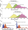
Fig 3. Dynamics of P. falciparum in malaria therapy patients.
(A) The first appearance of patent parasitemia (pale yellow) generally occurs at day 11 following an infectious bite, while gametocytemia (green) arises 11 days later because of the extended development and sequestration of P. falciparum gametocytes. Mosquitoes (pink) first become infected three days after the appearance of gametocytes. (B) The separation of timing of peaks in parasitemia (pale yellow), gametocytemia (green), and mosquito infection (pink) are similar to their first appearance but delayed by a week. (C) The period between the first appearance of patent parasitemia and gametocytemia (red) centers around 11 days, with some infections requiring as many as 20 days before patent gametocytemia after parasitemia has been observed. Mosquito infection is first observed in the initial days following patent gametocytemia (blue) but may not happen until weeks later. (D) The period between the peak of asexual parasitemia and gametocytemia (red) follows a similar appearance to the timing between initial appearances in C, but with more infections showing weeks between the peak in parasitemia and gametocytemia. The peak in mosquito infection (blue), however, occurs shortly after the peak in gametocytemia. n = 106. Patients without gametocytes or mosquito infection do not contribute to the time of appearance or peak of gametocytes and mosquito infection, respectively. Data in A and B are smoothed using a moving average including 5 consecutive days. (Data are courtesy of Dr. William E. Collins and Dr. Geoffrey M. Jeffery.)
Fig 4
Fig 4. Gametocyte sequestration in the bone marrow.
Several pathways may explain the enrichment of gametocytes in the extravascular compartment of the bone marrow and their subsequent release. (A) Asexual pRBCs adhere to the bone marrow endothelium and transmigrate into the bone marrow extravascular space. Once inside the bone marrow, asexual parasites either continue maturation producing asexual progeny or commit to production of gametocytes in the next cycle. (B) Sexually committed pRBCs specifically “home” to the bone marrow sinusoids by adhering to the bone marrow endothelium, after which they transmigrate into the extravascular space. Once in the extravascular compartment, the sexually committed pRBCs undergo schizogony resulting in the release of gametocyte-fated merozoites, which invade the abundant erythroid progenitor cells and for the most part develop attached to erythroblastic islands. (C) Similarly, sexually committed pRBCs could home to the bone marrow but not transmigrate into the extravascular space, perhaps because of their adhesion to the bone marrow endothelial cells or their low deformability. Upon schizont rupture within the bone marrow vasculature, the gametocyte-fated merozoites could enter the extravascular compartment and invade the erythroid progenitor cells. Merozoites from noncommitted asexual pRBCs may also enter the bone marrow and invade erythroid progenitor cells, either continuing asexual replication or forming gametocyte-fated merozoites the following cycle (not pictured). (D) Sexually committed pRBCs may not display a binding preference for bone marrow endothelial cells. Instead, sexually committed pRBCs may be formed in various asexual sequestration sites throughout the body with subsequent invasion of gametocyte-fated merozoites occurring in circulation, in a similar fashion to asexual invasion. Following intravascular invasion, however, the young gametocytes home to the bone marrow sinusoids and adhere to the endothelial cells, after which they transmigrate into the extravascular space. (E) Immature gametocytes display a markedly increased cellular rigidity [–81]. Upon maturation, however, deformability is rapidly restored, likely allowing the mature stage V gametocytes to exit the extravascular compartment and return to circulation, where they can be taken up by a feeding mosquito [–81].

References

    1. WHO (2014) WHO Malaria Report 2014. http://www.who.int/malaria/publications/world_malaria_report_2014/en/
    1. Lin JT, Saunders DL, Meshnick SR (2014) The role of submicroscopic parasitemia in malaria transmission: what is the evidence? Trends Parasitol 30: 183–190. 10.1016/j.pt.2014.02.004 - DOI - PMC - PubMed
    1. Lindblade KA, Steinhardt L, Samuels A, Kachur SP, Slutsker L (2013) The silent threat: asymptomatic parasitemia and malaria transmission. Expert Rev Anti Infect Ther 11: 623–639. 10.1586/eri.13.45 - DOI - PubMed
    1. Schneider P, Bousema T, Omar S, Gouagna L, Sawa P, et al. (2006) (Sub)microscopic Plasmodium falciparum gametocytaemia in Kenyan children after treatment with sulphadoxine-pyrimethamine monotherapy or in combination with artesunate. Int J Parasitol 36: 403–408. - PubMed
    1. Bousema T, Dinglasan RR, Morlais I, Gouagna LC, van Warmerdam T, et al. (2012) Mosquito feeding assays to determine the infectiousness of naturally infected Plasmodium falciparum gametocyte carriers. PLoS One 7: e42821 10.1371/journal.pone.0042821 - DOI - PMC - PubMed

Publication types