Review
. 2015 Jul;9(4):449-63.
doi: 10.5009/gnl14446.

The Pathogenesis and Management of Achalasia: Current Status and Future Directions

Affiliations
Review

The Pathogenesis and Management of Achalasia: Current Status and Future Directions

Fehmi Ates et al. Gut Liver. 2015 Jul.

Abstract

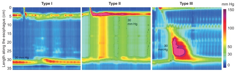
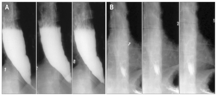
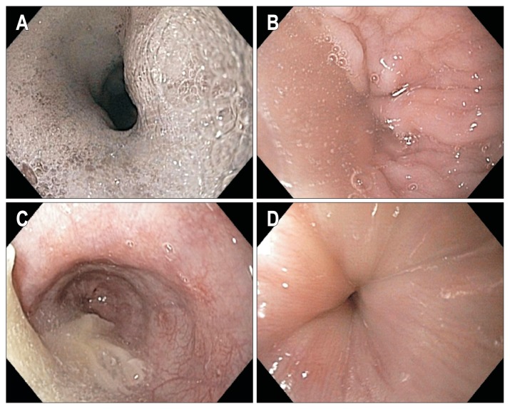
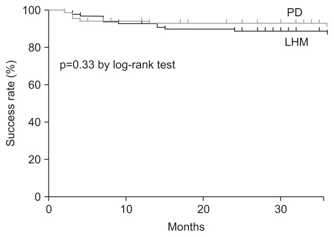
Achalasia is an esophageal motility disorder that is commonly misdiagnosed initially as gastroesophageal reflux disease. Patients with achalasia often complain of dysphagia with solids and liquids but may focus on regurgitation as the primary symptom, leading to initial misdiagnosis. Diagnostic tests for achalasia include esophageal motility testing, esophagogastroduodenoscopy and barium swallow. These tests play a complimentary role in establishing the diagnosis of suspected achalasia. High-resolution manometry has now identified three subtypes of achalasia, with therapeutic implications. Pneumatic dilation and surgical myotomy are the only definitive treatment options for patients with achalasia who can undergo surgery. Botulinum toxin injection into the lower esophageal sphincter should be reserved for those who cannot undergo definitive therapy. Close follow-up is paramount because many patients will have a recurrence of symptoms and require repeat treatment.

Keywords: Peroral esophageal myotomy; Pneumatic dilation; Surgical myotomy.


Figures

Fig. 1
Fig. 1
(A) Esophageal motor innervation by the vagus nerve; Auerbach’s and Meissner’s plexuses. (B) The striated muscle of the proximal esophagus is directly innervated by the somatic efferent cholinergic fibers of the vagus nerve originating from the nucleus ambiguus. In contrast, the smooth muscle of the distal esophagus is innervated by the preganglionic vagus nerve fibers from the dorsal motor nucleus. The preganglionic vagus fibers release acetylcholine, a neurotransmitter that affects two types of postganglionic neurons in the myenteric plexus, the excitatory cholinergic neurons and the inhibitory nitrinergic neurons. NO, nitric oxide; VIP, vasoactive intestinal polypeptide.
Fig. 2
Fig. 2
Both lower esophageal sphincter (LES) smooth muscle and the inhibitory neurons of the myenteric plexus have cholecystokinin receptors. (A) In a normal esophagus, administration of cholecystokinin-octapeptide (CCK-OP) results in LES relaxation because the inhibitory neurons override the direct excitation of the LES smooth muscle. (B) However, in achalasia, the LES smooth muscle excitation is unopposed due to the loss of the inhibitory neurons in the myenteric plexus. As a result, CCK-OP causes LES contraction.
Fig. 3
Fig. 3
In the early stage of achalasia, esophageal myenteric inflammation, caused by unknown host (genetic predisposition) and/or extrinsic (possibly viral) factors, may cause neuritis and ganglionitis with no ganglion cell loss or fibrosis. Functional esophageal dys-motility such as vigorous achalasia (type III achalasia) may be the predominant manifestation. Progressive destruction of the myenteric ganglion cells and neural fibrosis occurs, resulting in classic achalasia (types I and II).
Fig. 4
Fig. 4
Manometric tracings of achalasia by conventional water-perfused manometry: (A) simultaneous esophageal contractions associated with high lower esophageal sphincter (LES) pressure and (B) incomplete relaxation. High-resolution manometry tracings of (C) normal esophageal peristalsis and (D) achalasia showing simultaneous contractions along the esophagus with high E-sleeve LES pressure and incomplete relaxation. EGJ, esophagogastric junction; UES, upper esophageal sphincter.
Fig. 5
Fig. 5
High-resolution manometry of achalasia subtypes. Type I achalasia is associated with absent peristalsis and minimal esophageal body pressurization. Type II achalasia is associated with panesophageal pressurization related to a compression effect. Type III achalasia has evidence of abnormal contractility (spastic).
Fig. 6
Fig. 6
Timed barium swallow in achalasia. (A) Pretherapy retained barium in the esophagus at 1, 2, and 5 minutes after ingestion of barium. (B) Posttherapy barium swallow showing successful emptying of barium at all time intervals.
Fig. 7
Fig. 7
Endoscopic appearance of achalasia: (A) foam in the esophagus is often suggestive of poor motility and when combined with retained liquid (B) and food (C) along with a puckered gastroesophageal junction (D), should alert the endoscopist to the diagnosis of achalasia.
Fig. 8
Fig. 8
Kaplan-Meier graph showing equivalent success with pneumatic dilation (PD) versus laparoscopic Heller’s myotomy (LHM) over the study period.
Fig. 9
Fig. 9
Treatment algorithm for patients with achalasia. PD, pneumatic dilation.

References

    1. Sadowski DC, Ackah F, Jiang B, Svenson LW. Achalasia: incidence, prevalence and survival. A population-based study. Neurogastroenterol Motil. 2010;22:e256–e261. doi: 10.1111/j.1365-2982.2010.01511.x. - DOI - PubMed
    1. Gockel HR, Schumacher J, Gockel I, Lang H, Haaf T, Nöthen MM. Achalasia: will genetic studies provide insights? Hum Genet. 2010;128:353–364. doi: 10.1007/s00439-010-0874-8. - DOI - PubMed
    1. Vaezi MF, Richter JE. Diagnosis and management of achalasia: American College of Gastroenterology Practice Parameter Committee. Am J Gastroenterol. 1999;94:3406–3412. doi: 10.1111/j.1572-0241.1999.01639.x. - DOI - PubMed
    1. Francis DL, Katzka DA. Achalasia: update on the disease and its treatment. Gastroenterology. 2010;139:369–374. doi: 10.1053/j.gastro.2010.06.024. - DOI - PubMed
    1. Eckardt AJ, Eckardt VF. Treatment and surveillance strategies in achalasia: an update. Nat Rev Gastroenterol Hepatol. 2011;8:311–319. doi: 10.1038/nrgastro.2011.68. - DOI - PubMed

MeSH terms

LinkOut - more resources