Observational Study
. 2015 Aug;29(8):1099-110.
doi: 10.1038/eye.2015.103. Epub 2015 Jun 19.

In vivo detection of clinically non-apparent ocular surface inflammation in patients with meibomian gland dysfunction-associated refractory dry eye symptoms: a pilot study

Affiliations
Observational Study

In vivo detection of clinically non-apparent ocular surface inflammation in patients with meibomian gland dysfunction-associated refractory dry eye symptoms: a pilot study

Y Qazi et al. Eye (Lond). 2015 Aug.

Abstract

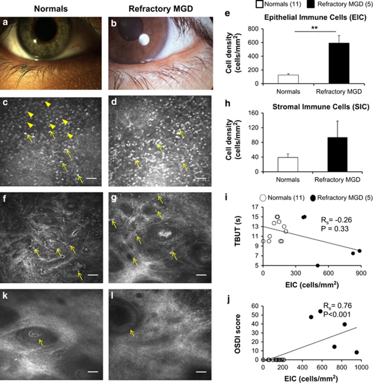
Purpose: The utility of in vivo confocal microscopy (IVCM) in the investigation of palpebral conjunctival and corneal inflammation in patients with meibomian gland dysfunction (MGD)-associated refractory dry eye symptoms following gland expression, despite objective clinical improvement.

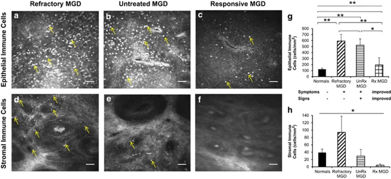
Methods: A retrospective, observational pilot study was conducted evaluating five patients with MGD-associated refractory dry eye symptoms and three control groups: symptomatic untreated MGD patients (n=3), treatment-responsive MGD patients with improved symptoms (n=3) and asymptomatic healthy normals (n=11). Ocular surface disease index (OSDI) scores, tear break-up time (TBUT), the number of meibomian glands yielding liquid secretion (MGYLS), palpebral conjunctival epithelial and substantia propria immune cell (EIC, SIC), and corneal dendritic cell (DC) densities were measured.

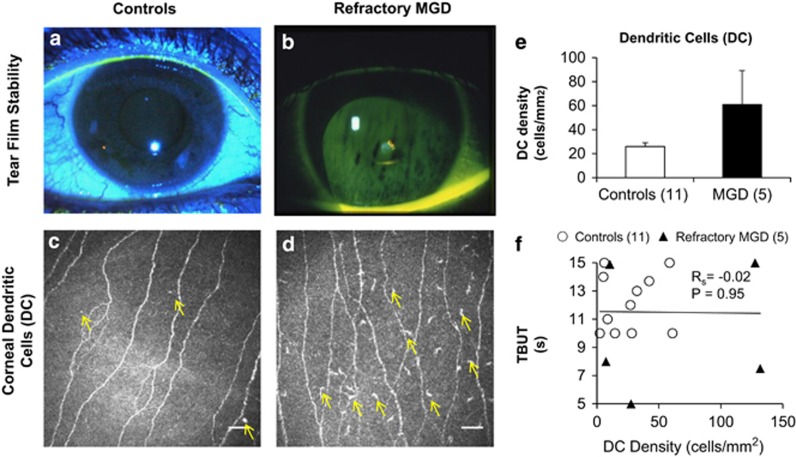
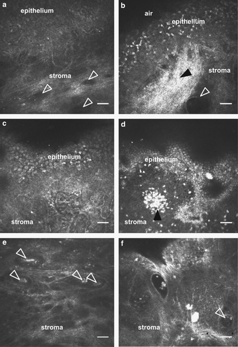
Results: Despite clinical improvement (TBUT: 6.4±1.2 s to 10.1±2.1 s, P=0.03; MGYLS: 3.5±0.8 glands to 7.0±1.1 glands, P=0.13) and a normal clinical examination post treatment, MGD patients remained symptomatic. IVCM revealed increased immune cells in the palpebral conjunctiva (refractory MGD EIC=592.6±110.1 cells/mm2 untreated MGD EIC=522.6±104.7 cells/mm2, P=0.69; responsive MGD EIC=194.9±119.4 cells/mm2, P<0.01; normals EIC=123.7±19.2 cells/mm2, P< 0.001), but not the cornea (refractory MGD DC=60.9±28.3 cells/mm2; normals DC=25.9±6.3 cells/mm2; P=0.43). EIC did not correlate with TBUT (Rs=-0.26, P=0.33). OSDI scores correlated with both EIC (Rs=0.76, P<0.001) and TBUT (Rs=-0.69, P<0.01) but not SIC. Intraglandular immune cells were also seen.

Conclusion: MGD-associated refractory symptoms and the symptom-sign disparity may be explained by clinically non-apparent, active inflammation of the palpebral conjunctiva as detected by IVCM. These patients may benefit from anti-inflammatory therapy.


Figures

Figure 1
Figure 1
Comparison of refractory MGD patients with healthy normal controls on in vivo confocal microscopy. Representative slit-lamp photographs of the eyelid margin in healthy eyes (a) and patients with MGD (b), showing plugging and pouting of meibomian glands in MGD (b). As compared with eyes of healthy, normal, asymptomatic controls (c, f; arrows) with numerous goblet cells (c, arrowheads), en face corneal in vivo confocal micrographs (HRT 3/RCM, Heidelberg Engineering, Germany) of MGD patients with refractory symptoms demonstrated increased infiltration of immune cells (d, g; arrows) in both the conjunctival epithelium (EIC=592.6±110.1 cells/mm2 vs 123.7±19.2 cells/mm2 P<0.01; d,e) and stroma (SIC=93.9±44.2 cells/mm2 vs 38.8±9.5 cells/mm2, P=0.11; g,h). The near five-fold increase in conjunctival epithelial immune cell density (EIC; e) showed a trend of inverse correlation with tear break-up time (TBUT; Rs=−0.26, P=0.33, n=16; i), and a highly significant positive correlation with symptom severity (OSDI scores; Rs=0.76, P<0.001, n=16; j) supporting the relationship between symptom severity in MGD and density of palpebral conjunctival EICs. In some healthy eyes, occasional intraglandular immune cells within ductules of presumed meibomian glands were observed (k, arrow) without ductular dilatation (lumen diameter<50 μm). Dilated glands can be seen in patients with refractory MGD-associated dry eye symptoms (l, arrow), suggestive of occult deep-seated glandular obstruction. Results are reported as mean±SEM. A probability value (P) of less than 0.05 was considered statistically significant (*), whereas P<0.01 was considered highly statistically significant (**). Legend: numbers in parentheses represent the number of patients per group. Scale bar represents 40 μm.
Figure 2
Figure 2
Comparison of refractory MGD, untreated symptomatic MGD, and treatment-responsive MGD patients on in vivo confocal microscopy. En face in vivo confocal micrographs (HRT 3/RCM, Heidelberg Engineering, Germany) of the palpebral conjunctiva showed increased infiltration of immune cells in both the conjunctival epithelium (a–c, g; analysis of variance (ANOVA), P<0.0001) and stroma (d–f, h; ANOVA, P=0.03) of eyes with MGD-associated refractory dry eye symptoms. Epithelial immune cell density (EIC) in refractory patients (592.6±110.1 cells/mm2; a, g) was nearly five-fold greater than normals (EIC=123.7±19.2 cells/mm2 P<0.01; g), comparable to epithelial inflammation in untreated, highly symptomatic MGD patients (522.6±104.7 cells/mm2, P=0.69; b, g), and three-fold higher than that of treatment-responsive and less symptomatic MGD patients (194.9±119.4 cells/mm2, P<0.01; c, g). Stromal immune cell density (SIC) was increased several-fold in refractory patients (93.9±44.2 cells/mm2; d, h) as compared with untreated, symptomatic MGD patients (29.2±18.4 cells/mm2, P=0.33; e, h), and treatment-responsive, less symptomatic MGD patients (4.6±3.1 cells/mm2, P=0.18; f, h). Results are reported as mean±SEM. A probability value (P) of less than 0.05 was considered statistically significant (*), whereas P of less than 0.01 was considered highly statistically significant (**). Numbers in parentheses represent the number of patients per group: normals (11), refractory MGD (5), unRx MGD (3), Rx MGD (3). Axes: untreated (UnRx), treatment-responsive (Rx).
Figure 3
Figure 3
Corneal dendritic cells (DCs) in patients with persistent meibomian gland dysfunction (MGD)-associated dry eye symptoms. Representative slit-lamp photographs of tear film stability in healthy asymptomatic eyes (a) and patients with refractory MGD (b). As compared with healthy eyes (c, e; arrows indicate DCs), corneal in vivo confocal micrographs demonstrate no significant difference (P=0.11) in the corneal DC densities of patients with refractory symptoms (d, e; arrows indicate DCs). Although there is a trend of increased corneal DC density in patients with persistent dry eye symptoms (c, d, e), the difference is neither statistically significant (P=0.43) nor does it bear correlation with tear break-up time (TBUT; (f), Rs=−0.02, P=0.95). Results are reported as mean±SEM. A probability value (P) of less than 0.05 is considered statistically significant. The numbers in parentheses represent the number of patients per group. Scale bar represents 40 μm.
Figure 4
Figure 4
Qualitative features of the palpebral conjunctiva and associated glands in MGD-associated persistent dry eye symptoms using in vivo confocal microscopy (IVCM). Compared with the palpebral conjunctiva of healthy asymptomatic controls (a, c, e), IVCM micrographs of MGD patients with persistent symptoms (b, d, f) showed sub-epithelial fibrosis (b, filled arrowhead), dilated stromal glands with cellular content (b, f empty arrowhead), conjunctiva-associated lymphoid tissue (d, filled arrowhead). Some intrastromal glands with cellular content (f, arrowhead) were surrounded by visible stromal blood vessels (f, asterisks). These intrastromal glandular structures with intraglandular content were seen at a depth of ~200 μm from the surface of palpebral conjunctival epithelium, suggestive of possible meibomian glands. Scale bar represents 40 μm.

References

    1. Viso E, Gude F, Rodriguez-Ares MT. The association of meibomian gland dysfunction and other common ocular diseases with dry eye: a population-based study in Spain. Cornea. 2011;30 (1:1–6. - PubMed
    1. Bron AJ, Tiffany JM. The contribution of meibomian disease to dry eye. Ocul Surf. 2004;2 (2:149–165. - PubMed
    1. Rege A, Kulkarni V, Puthran N, Khandgave T. A clinical study of subtype-based prevalence of dry eye. J Clin Diagn Res. 2013;7 (10:2207–2210. - PMC - PubMed
    1. Nichols KK. The international workshop on meibomian gland dysfunction: introduction. Invest Ophthalmol Vis Sci. 2011;52 (4:1917–1921. - PMC - PubMed
    1. Schaumberg DA, Nichols JJ, Papas EB, Tong L, Uchino M, Nichols KK. The international workshop on meibomian gland dysfunction: report of the subcommittee on the epidemiology of, and associated risk factors for, MGD. Invest Ophthalmol Vis Sci. 2011;52 (4:1994–2005. - PMC - PubMed

Publication types

LinkOut - more resources