

Osteonecrosis of the femoral head: diagnosis and classification systems
- PMID: 26088795
- PMCID: PMC4596207
- DOI: 10.1007/s12178-015-9278-7
Osteonecrosis of the femoral head: diagnosis and classification systems
Abstract
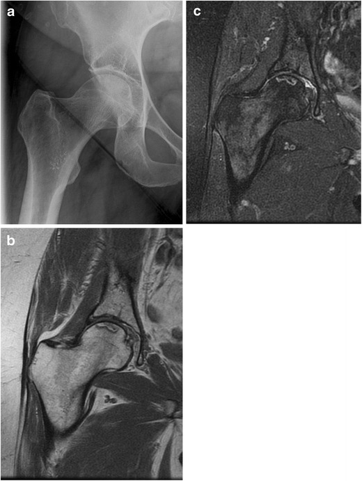
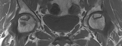
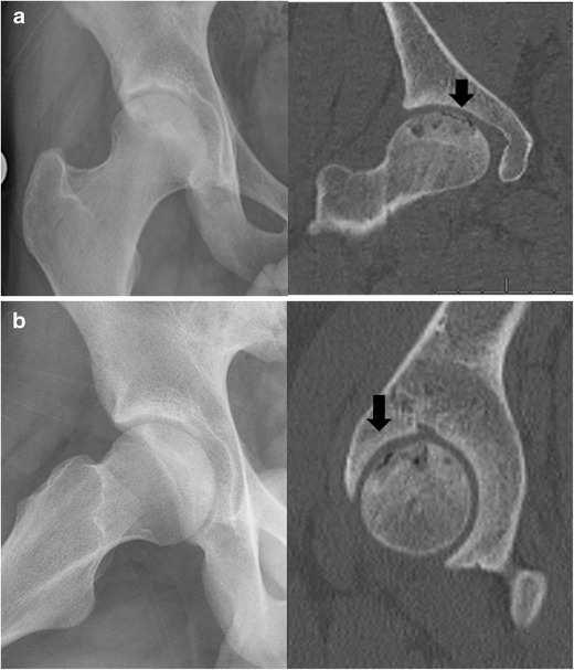
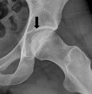
Osteonecrosis of femoral head is a rare but disabling condition that usually results in progressive femoral head collapse and secondary arthritis necessitating total hip arthroplasty if not treated appropriately in early stages. However, early diagnosis is challenging as the onset of disease is insidious and the symptoms and signs are usually minimal and nonspecific until it becomes advanced. Of several diagnostic modalities, magnetic resonance imaging (MRI) is considered the imaging method of choice with the highest sensitivity and specificity, while detection of potential risk factors is very important as well. Many investigators have developed several different classification systems; however, there still is controversy regarding the optimal classification system. Diagnostic methods and the evolution of different classification systems will be reviewed in this paper.
Figures
References
-
- Lieberman JR, Berry DJ, Mont MA, Aaron RK, Callaghan JJ, Rajadhyaksha AD, et al. Osteonecrosis of the hip: management in the 21st century. Instr Course Lect. 2003;52:337–55. - PubMed
-
- Steinberg ME, Steinberg DR. Osteonecrosis: Historical perspective. In: Koo KH, Mont MA, Jones LC, editors. Osteonecrosis. Heidelberg: Springer; 2014. p. 3–15. The study describes historical perspective of diagnosis, classification, and treatment of osteonecrosis.
-
- Lu-Yao GL, Keller RB, Littenberg B, Wennberg JE. Outcomes after displaced fractures of the femoral neck. A meta-analysis of one hundred and six published reports. J Bone Joint Surg Am. 1994;76(1):15–25. - PubMed
-
- Min BW, Kim SJ. Avascular necrosis of the femoral head after osteosynthesis of femoral neck fracture. Orthopedics. 2011;34(5):349. - PubMed
LinkOut - more resources
Full Text Sources
Other Literature Sources
