. 2015 Jun 19;348(6241):1361-5.
doi: 10.1126/science.aaa5264.

SIGNAL TRANSDUCTION. Structural basis for nucleotide exchange in heterotrimeric G proteins

Affiliations

SIGNAL TRANSDUCTION. Structural basis for nucleotide exchange in heterotrimeric G proteins

Ron O Dror et al. Science. .

Abstract

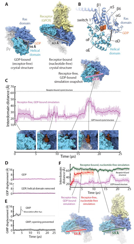
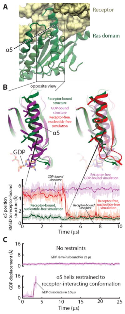
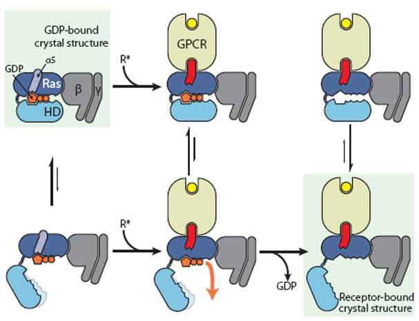
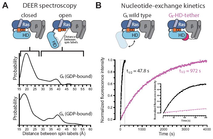
G protein-coupled receptors (GPCRs) relay diverse extracellular signals into cells by catalyzing nucleotide release from heterotrimeric G proteins, but the mechanism underlying this quintessential molecular signaling event has remained unclear. Here we use atomic-level simulations to elucidate the nucleotide-release mechanism. We find that the G protein α subunit Ras and helical domains-previously observed to separate widely upon receptor binding to expose the nucleotide-binding site-separate spontaneously and frequently even in the absence of a receptor. Domain separation is necessary but not sufficient for rapid nucleotide release. Rather, receptors catalyze nucleotide release by favoring an internal structural rearrangement of the Ras domain that weakens its nucleotide affinity. We use double electron-electron resonance spectroscopy and protein engineering to confirm predictions of our computationally determined mechanism.


Figures

Figure 1
Figure 1
The Ras and helical domains of the G protein α subunit separate spontaneously and frequently when GDP is bound, even in the absence of a receptor. (A) The Ras and helical domains are tightly apposed in all nucleotide-bound G protein crystal structures, enveloping the nucleotide (left: GDP-bound Gt heterotrimer; PDB entry 1GOT), but are dramatically separated in the receptor-bound, nucleotide-free structure (right: β2-adrenergic receptor–Gs heterotrimer [β2AR–Gs] complex; PDB entry 3SN6). GDP is colored orange, the Ras domain blue, the helical domain cyan, Gβγ gray, and the receptor yellow. The degree of domain separation is represented by a thick black line connecting Ala134 and Glu272 in Gαt or the corresponding Ala161 and Glu299 in Gαs, with both ends connected by white lines to a pivot point near Thr166 (Gαt) or Ser193 (Gαs). (B) Key structural motifs of the α subunit, illustrated using the GDP-bound Gt structure. (C) Spontaneous domain separation provides an exit pathway for GDP. In simulations of receptor-free, GDP-bound Gt, the Ala134–Glu272 distance varies substantially as the domains fluctuate between apposed and separated conformations; raw (light purple) and smoothed (250-ns moving average; dark purple) data are shown. Representative molecular simulation snapshots (top: overview; bottom: nucleotide-binding site) display varying degrees of GDP exposure. Data are from simulation 2 (Table S1). (D) Domain separation is not sufficient for rapid nucleotide release. GDP remains tightly bound to receptor-free Gt (top), even with the helical domain removed (bottom; traces show displacement of the centroid of the nucleotide non-hydrogen atoms relative to the crystal structure). Data from simulations 2 and 33. (E) Domain separation is necessary for rapid nucleotide release. GMP dissociates spontaneously from receptor-free Gt (top) but remains bound when the interdomain distance is artificially restrained to prevent domain separation (bottom). Data from simulations 16 and 31. (F) Domain separation is greater in the absence of a nucleotide. In simulations initiated from the receptor-free, GDP-bound Gt crystal structure, but with the GDP removed, the Ras and helical domains exhibited extensive and prolonged separation (red trace; left-hand snapshot). In simulations of the β2AR–Gs complex, also nucleotide-free, the helical domain remained widely separated from the Ras domain, although it typically moved away from the membrane toward the beta propeller of Gβγ (green; right-hand snapshot). GDP-bound Gt simulation data from panel C are replicated for reference (purple). See SM for details on structural renderings. Data from simulations 2, 14, and 22.
Figure 2
Figure 2
Receptor-induced displacement of the Gα C-terminal α5 helix disrupts key GDP contacts, thereby promoting nucleotide release. (A) In the receptor-bound, nucleotide-free crystal structure (PDB entry 3SN6) crystal structure, α5 docks into receptor. (B) (Top, left) Superimposition of receptor-free, GDP-bound (PDB entry 1GOT; purple) and receptor-bound, nucleotide-free (PDB entry 3SN6; green) crystal structures shows the displacement of α5, relative to the rest of the Ras domain, that occurs when a G protein binds to an activated receptor. (Top, right) In a simulation initialized from a receptor-free, GDP-bound Gt structure but with GDP removed (red), α5 spontaneously rotated 60° and translated 5 Å, adopting a position distal from the nucleotide-binding site that closely matched that of the β2AR–Gs complex (green). Several side chains in the α5 helix and α5–β6 loop are shown to facilitate comparison between structures. (Bottom) The position of α5 in this simulation (red) changed abruptly at ~4.5 μs to match that of the β2AR–Gs complex; the α5 position was stable in simulations of receptor-free, GDP-bound Gt (purple) and of the β2AR–Gs complex (green). Data are from simulations 5, 12, and 22 (Table S1). (C) Forcing α5 into the distal conformation accelerates nucleotide release in simulation. Temperature-accelerated MD simulations allow observation of GDP release on computationally accessible timescales, but only when α5 is restrained to the distal conformation (i.e., the conformation observed in the β2AR–Gs complex). Receptor-free, GDP-bound Gt was simulated without (top) or with (bottom) restraints on α5 (see SOM). GDP displacement is measured as in Fig. 1. Data are from simulations 55 and 56.
Figure 3
Figure 3
Proposed mechanism of receptor-catalyzed nucleotide release. (Left) The Ras and helical domains (Ras and HD) separate frequently, even in the absence of a receptor, but such separation does not usually lead to GDP release. This rapid (relative to overall GDP release) equilibrium favors the closed state (top). (Middle) Binding of an activated receptor induces a Ras domain conformational change—displacement of α5 away from GDP—that weakens interactions between GDP and the Ras domain, allowing GDP to escape when the Gα domains happen to spontaneously separate (bottom). (Right) Loss of GDP shifts the equilibrium toward Gα conformations with widely separated domains (bottom).
Figure 4
Figure 4
Experimental validation of spontaneous Gα domain separation in GDP-bound heterotrimeric G proteins and its role in nucleotide exchange. (A) DEER distance distributions measured between spin labels attached to the Ras and helical domains of Gi (Glu238 and Arg90) and Gs (Asn261 and Asn112) show multiple distance peaks, consistent with an equilibrium between closed and open conformations of the α subunit in the presence of GDP, despite the absence of an activated receptor. These distance distributions extend to much larger values than would be expected if the G proteins maintained their crystallographic nucleotide-bound conformations (Fig. S15). (B) Domain separation impacts the basal GDP release rate. The Gi-HD-tether construct (Fig. S16), designed to restrict domain separation, exchanges nucleotides 20-fold more slowly than Gi wild type, under conditions where GDP release is rate-limiting. GDP release was monitored by BODIPY-GTPγS binding kinetics, shown for Gi wild type (black) and Gi-HD-tether (purple). The inset corresponds to the gray dashed box.

References

    1. Oldham WM, Hamm HE. Heterotrimeric G protein activation by G-protein-coupled receptors. Nat Rev Mol Cell Biol. 2008;9:60–71. - PubMed
    1. Sprang SR. G protein mechanisms: Insights from structural analysis. Ann Rev Biochem. 1997;66:639–678. - PubMed
    1. Rosenbaum DM, Rasmussen SGF, Kobilka BK. The structure and function of G-protein-coupled receptors. Nature. 2009;459:356–363. - PMC - PubMed
    1. Rasmussen SGF, et al. Crystal structure of the β2 adrenergic receptor–Gs protein complex. Nature. 2011;477:549–555. - PMC - PubMed
    1. Venkatakrishnan AJ, Deupi X, Lebon G, Tate CG, Schertler GF, Babu MM. Molecular signatures of G-protein-coupled receptors. Nature. 2013;494:185–194. - PubMed

Publication types

MeSH terms

Substances