. 2015 Jun 3:9:196.
doi: 10.3389/fnins.2015.00196. eCollection 2015.

Loss of tau rescues inflammation-mediated neurodegeneration

Affiliations

Loss of tau rescues inflammation-mediated neurodegeneration

Nicole Maphis et al. Front Neurosci. .

Abstract

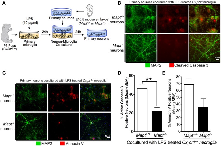
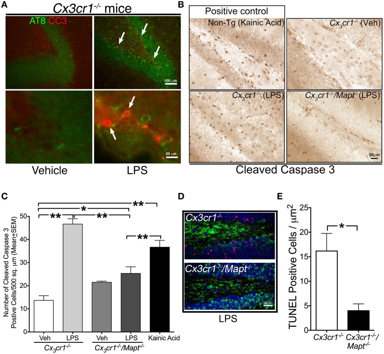
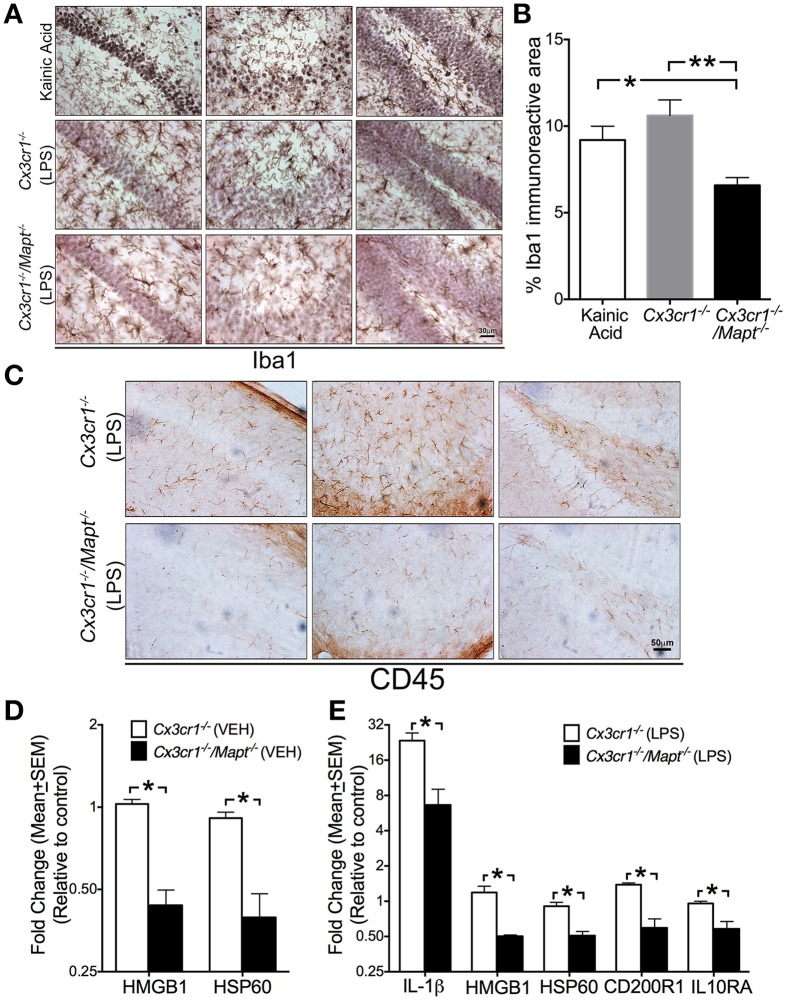
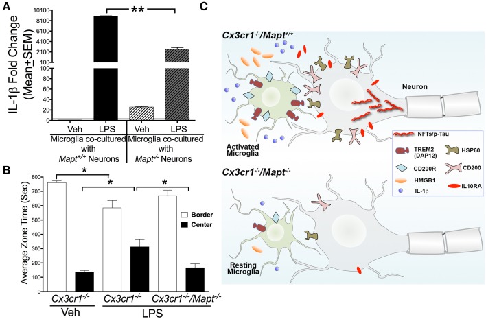
Neuroinflammation is one of the neuropathological hallmarks of Alzheimer's disease (AD) and related tauopathies. Activated microglia spatially coexist with microtubule-associated protein tau (Mapt or tau)-burdened neurons in the brains of human AD and non-AD tauopathies. Numerous studies have suggested that neuroinflammation precedes tau pathology and that induction or blockage of neuroinflammation via lipopolysaccharide (LPS) or anti-inflammatory compounds (such as FK506) accelerate or block tau pathology, respectively in several animal models of tauopathy. We have previously demonstrated that microglia-mediated neuroinflammation via deficiency of the microglia-specific chemokine (fractalkine) receptor, CX3CR1, promotes tau pathology and neurodegeneration in a mouse model of LPS-induced systemic inflammation. Here, we demonstrate that tau mediates the neurotoxic effects of LPS in Cx3cr1 (-/-) mice. First, Mapt (+/+) neurons displayed elevated levels of Annexin V (A5) and TUNEL (markers of neurodegeneration) when co-cultured with LPS-treated Cx3cr1 (-/-)microglia, which is rescued in Mapt (-/-) neurons. Second, a neuronal population positive for phospho-S199 (AT8) tau in the dentate gyrus is also positive for activated or cleaved caspase (CC3) in the LPS-treated Cx3cr1 (-/-) mice. Third, genetic deficiency for tau in Cx3cr1 (-/-) mice resulted in reduced microglial activation, altered expression of inflammatory genes and a significant reduction in the number of neurons positive for CC3 compared to Cx3cr1 (-/-)mice. Finally, Cx3cr1 (-/-)mice exposed to LPS displayed a lack of inhibition in an open field exploratory behavioral test, which is rescued by tau deficiency. Taken together, our results suggest that pathological alterations in tau mediate inflammation-induced neurotoxicity and that deficiency of Mapt is neuroprotective. Thus, therapeutic approaches toward either reducing tau levels or blocking neuroinflammatory pathways may serve as a potential strategy in treating tauopathies.

Keywords: Alzheimer's disease; CX3CR1; microglia; microtubule associated protein tau (MAPT); neurodegeneration; neuroinflammation; tau protein; tauopathies.


Figures

Figure 1
Figure 1
Loss of tau reduces inflammation-induced expression of CC3 and Annexin V (A5) in primary neurons. (A) Schematic showing the neuron-microglia co-culture experiments. Primary cortical neurons at 21 days in vitro (DIV) (Mapt+/+ or Mapt−/−) were co-cultured with LPS (1 μg/ml; 24 h)-stimulated Cx3cr1−/− microglia for 24 h. The neurons were fixed and stained for CC3 and A5. (B,C) MAP2 positive Mapt−/− neurons show reduced CC3 and A5 staining compared to Mapt+/+ neurons. The blue label in the last panel shows DAPI. Scale bar 10 μm. (D) The percentage of CC3+MAP2+ and A5+MAP2+ neurons revealed a statistically significant (**p < 0.01 for CC3; unpaired t-test; n = 3 independent experiments; mean ± SEM) reduction for CC3+MAP2+ for Mapt−/− neurons compared to Mapt+/+ neurons. (E) Quantification of A5+MAP2+ neurons reveals trend toward reduced A5+MAP2+ immunoreactivity in Mapt−/− neurons compared to Mapt+/+ neurons.
Figure 2
Figure 2
Tau deficiency reduces LPS-induced neurotoxicity in Cx3cr1−/− mice. (A) Two-month old Cx3cr1−/− mice were injected with single dose of vehicle (left panel) or LPS (right panel) (1 mg/kg b.w, daily for 3 days; i.p). Brain sections were double immune-labeled with AT8 (phospho-tau at Ser199) and CC3 antibodies. Presence of AT8 (green) and CC3 (red, arrows) positive cells were detected only in LPS treated samples, primarily in the same sub-population of dentate gyrus. Top panel-low magnification; bottom panel-high magnification. Scale bar 100 μm in top panels and 20 μm in bottom panels. (B) The number of cells immunoreactive for CC3 is relatively higher in the DG of 2-month-old Cx3cr1−/− mice administered with LPS compared to 2-month-old Cx3cr1−/− mice injected with vehicle (Veh). Deficiency of tau in Cx3cr1−/−/Mapt−/− mice shows markedly reduced numbers of CC3+ cells following administration of a similar concentration of LPS. As a positive control, 2-month-old non-transgenic mice treated with kainic acid (20 mg/kg b.w, single dose; i.p.) showed a substantial number of CC3+ cells in the DG. Scale 30 μm. (C) Quantification of CC3+ cells reveals a statistically significant (**p < 0.01; One-Way ANOVA with Tukey multiple comparison test; n = 4 mice per group) increase in CC3+ cells in the LPS treated Cx3cr1−/− mice compared to the Veh treated mice. Note LPS-treated Cx3cr1−/−/Mapt−/− mice showed significantly less CC3+ cells (**p < 0.01; One-Way ANOVA with Tukey multiple comparison test; n = 4; mean ± SEM) compared to LPS-treated Cx3cr1−/− mice or KA-treated mice. Other comparisons are also shown (*p < 0.05 or **p < 0.01). (D,E) The number of NeuN+ neurons (green), which are immunoreactive for TUNEL (red) in the granule cell layer of DG is significantly (*p < 0.05; unpaired t-test; n = 4 mice per genotype; mean ± SEM) higher in the LPS-treated Cx3cr1−/− mice compared to the LPS treated Cx3cr1−/−/Mapt−/− mice. Scale bar is 50 μm.
Figure 3
Figure 3
Tau deficiency in neurons alters microglial immune response. (A,B) Iba1+ microglia display activated morphology in the CA1 (left column in A), CA3 (middle column in A) and dentate gyrus (right column in A) of 2-month-old C57BL/6 injected with KA (20 mg/kg b.w, single dose; i.p) and in 2-month-old Cx3cr1−/− mice administered with LPS (1 mg/kg b.w, daily for 3 days; i.p). Note that the 2-month-old Cx3cr1−/−/Mapt−/− mice administered with the same dose of LPS show relatively lower numbers of Iba1+ microglia, which also appear less activated. Morphometric quantification reveals a statistically (*p < 0.05 vs. KA treated mice; **p < 0.01 vs. LPS treated Cx3cr1−/− mice; One-Way ANOVA with Tukey multiple comparison test; n = 4 mice/genotype; mean ± SEM) significant decrease in the percentage of Iba1+ area in the CA1 subfield in the LPS treated Cx3cr1−/−/Mapt−/− mice compared to LPS treated Cx3cr1−/− mice or KA treated controls. Scale 30 μm. (C) Reduced CD45+ cells in the CA1, CA3, and DG subfields in LPS treated Cx3cr1−/−/Mapt−/− mice compared to LPS treated Cx3cr1−/− mice. Scale bar 50 μm. (D,E) Quantitative real-time PCR (qRT-PCR) analysis for HMGB1 and HSP60 transcripts (in the vehicle-treated groups; in D) and IL-1β, HMGB1, HSP60, CD200R1, and IL10RA (in the LPS-treated groups; in E) revealed a significant decrease (normalized to non-transgenics; *p < 0.05; One-Way ANOVA followed by Tukey post-hoc test; n = 4 mice per group; mean ± SEM) in the inflammatory molecules in the hemibrains of 2-month-old Cx3cr1−/−/Mapt−/− mice compared to Cx3cr1−/−mice.
Figure 4
Figure 4
Tau deficiency reduces microglial-IL-1β secretion in vitro and lack of inhibition in the Cx3cr1−/− mice following LPS administration. (A) qRT-PCR analysis reveal LPS induced pro-IL-1β expression in Cx3cr1−/− microglia co-cultured with Mapt+/+ neurons is significantly (**p < 0.01; One-Way ANOVA with Tukey multiple comparison test; n = 3 independent co-cultures; mean ± SEM) higher compared to Cx3cr1−/− microglia co-cultured with Mapt−/− neurons. (B) Two-month-old Cx3cr1−/− and Cx3cr1−/−/Mapt−/− mice administered with LPS (1 mg/kg b.w, daily for 3 days; i.p) or vehicle were subjected to open-field behavioral test (for 15 min) after 7 days post-LPS treatment. LPS treated Cx3cr1−/− mice spent significantly (*p < 0.05; One-Way ANOVA with Tukey multiple comparison test; n = 4 mice per genotype/group; mean ± SEM) less time in the border, but more time in the center of the maze, compared to vehicle injected Cx3cr1−/− mice or LPS injected Cx3cr1−/−/Mapt−/− mice. (C) The working model suggests that in Cx3cr1−/−/Mapt+/+ mice, LPS induces hyperphosphorylation (p-Tau) and possible aggregation of tau as neurofibrillary tangles (NFTs) due in part to the sustained activation of Cx3cr1−/− microglia. This results in upregulation of various inflammatory regulators including neuronally derived ligands (HSP60, CD200, HMGB1), microglial receptors (TREM2/DAP12 and CD200R), IL-1β and IL10RA that are needed to restrain reactive microglia. Despite activation of various negative regulatory pathways of microglia, microglia continue to show significant activation due to the deficiency of the CX3CL1-CX3CR1 communication and LPS. Sustained activation of Cx3cr1−/− microglia leads to neurotoxicity possibly mediated by further tau hyperphosphorylation and by the elevated IL-1β signaling. Deficiency of tau in Cx3cr1−/−/Mapt−/− mice show reduced microglial activation, significant reduction in inflammatory mediators including IL-1β and HMGB1, overall decrease in neuron-microglia restraint signaling molecules and thus prevents inflammation-induced, tau-mediated, neurotoxicity.

References

    1. Abbondante S., Baglietto-Vargas D., Rodriguez-Ortiz C. J., Estrada-Hernandez T., Medeiros R., Laferla F. M. (2014). Genetic ablation of tau mitigates cognitive impairment induced by type 1 diabetes. Am. J. Pathol. 184, 819–826. 10.1016/j.ajpath.2013.11.021 - DOI - PMC - PubMed
    1. Ahmed T., van der Jeugd A., Blum D., Galas M. C., D'hooge R., Buee L., et al. . (2014). Cognition and hippocampal synaptic plasticity in mice with a homozygous tau deletion. Neurobiol. Aging 35, 2474–2478. 10.1016/j.neurobiolaging.2014.05.005 - DOI - PubMed
    1. Akiyama H., Arai T., Kondo H., Tanno E., Haga C., Ikeda K. (2000). Cell mediators of inflammation in the Alzheimer disease brain. Alzheimer. Dis. Assoc. Disord. 14(Suppl. 1), S47–S53. 10.1097/00002093-200000001-00008 - DOI - PubMed
    1. Barclay A. N., Wright G. J., Brooke G., Brown M. H. (2002). CD200 and membrane protein interactions in the control of myeloid cells. Trends Immunol. 23, 285–290. 10.1016/S1471-4906(02)02223-8 - DOI - PubMed
    1. Bazan J. F., Bacon K. B., Hardiman G., Wang W., Soo K., Rossi D., et al. . (1997). A new class of membrane-bound chemokine with a CX3C motif. Nature 385, 640–644. 10.1038/385640a0 - DOI - PubMed