. 2015;11(8):1389-407.
doi: 10.1080/15548627.2015.1063769.

Coxsackievirus can exploit LC3 in both autophagy-dependent and -independent manners in vivo

Affiliations

Coxsackievirus can exploit LC3 in both autophagy-dependent and -independent manners in vivo

Mehrdad Alirezaei et al. Autophagy. 2015.

Abstract

RNA viruses modify intracellular membranes to produce replication scaffolds. In pancreatic cells, coxsackievirus B3 (CVB3) hijacks membranes from the autophagy pathway, and in vivo disruption of acinar cell autophagy dramatically delays CVB3 replication. This is reversed by expression of GFP-LC3, indicating that CVB3 may acquire membranes from an alternative, autophagy-independent, source(s). Herein, using 3 recombinant CVB3s (rCVB3s) encoding different proteins (proLC3, proLC3(G120A), or ATG4B(C74A)), we show that CVB3 is, indeed, flexible in its utilization of cellular membranes. When compared with a control rCVB3, all 3 viruses replicated to high titers in vivo, and caused severe pancreatitis. Most importantly, each virus appeared to subvert membranes in a unique manner. The proLC3 virus produced a large quantity of LC3-I which binds to phosphatidylethanolamine (PE), affording access to the autophagy pathway. The proLC3(G120A) protein cannot attach to PE, and instead binds to the ER-resident protein SEL1L, potentially providing an autophagy-independent source of membranes. Finally, the ATG4B(C74A) protein sequestered host cell LC3-I, causing accumulation of immature phagophores, and massive membrane rearrangement. Taken together, our data indicate that some RNA viruses can exploit a variety of different intracellular membranes, potentially maximizing their replication in each of the diverse cell types that they infect in vivo.

Keywords: ER; ERAD; LC3; RNA virus; SEL1L; autophagy; coxsackievirus; enterovirus; membranes; pancreas.


Figures
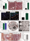
Figure 1.
Figure 1.
For figure legend, see page 1392.
Figure 2.
Figure 2.
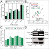
Design and characterization of 3 rCVB3s to dissect the impact of various forms of LC3. (A) A schematic summary of proLC3 processing into LC3-I and LC3-II is shown, and includes the relevant amino acid residue at the Ct of the protein (see text). The Ct residues of the mutated form, proLC3G120A, is shown at the top-right. (B) One-step growth curves of rCVB3s and WTCVB3 are shown (HeLa cells, MOI = 10). (C) Protein was harvested from HeLa cells (8 h p.i.), and the expression of the viral protein VP1, and of GAPDH control, were assessed by western blot. (D) The samples were applied on a separate gel, this time using differing loading amounts, for reasons explained in the text. The relative loading amounts can be determined from the GAPDH data (lower blot). For all viruses, the quantities of autophagy-related products were determined (upper blot) and, for the Atg4BC74A virus, the amount of ATG4BC74A expression was evaluated (middle blot). Control samples were included from uninfected mouse embryonic fibroblasts (MEF), some of which had been incubated with the late autophagy inhibitor bafilomycin A1 (Baf). (E) Equal quantities of protein from cells infected either with proLC3-CVB3, or proLC3G120A-CVB3, were applied to a polyacrylamide gel, and for each, the gel region containing proteins of 12 to 18 KDa was excised. Proteins were extracted, and analyzed using nano LC-MS/MS. The predicted protein sequences from the N termini of the recombinant polyproteins are shown. Native viral amino-acids are in lower case, and recombinant (proLC3 or proLC3G120A) residues are in upper case. Red font indicates peptides that were identified by nano LC-MS/MS; the uncleavable FATAMAV sequence in the proLC3G120A protein (see text) is underlined. (F) Summary of the observed autophagy-related changes for each of the 3 new rCVB3s.
Figure 4.
Figure 4.
Ultrastructural changes in cells infected with rCVB3s expressing autophagy-related proteins. Acinar cells isolated from pancreata of WT mice were infected (MOI 100) with each of 3 indicated rCVB3s: proLC3-CVB3 (A-C), proLC3G120A-CVB3 (D-F) or Atg4BC74-CVB3 (G-I). Eighteen h later, the cells were evaluated by transmission electron microscopy. n = 80 acinar cells infected with proLC3-CVB3; n = 85 acinar cells infected with proLC3G120A-CVB3; and n = 92 acinar cells infected with Atg4BC74A-CVB3 from 3 independent experiments. For each of the 3 different virus-infected cell types, the diameters of numerous DMVs (38 to 90) were measured; there was no significant difference in the sizes of the DMVs. Scale bars are included for all panels. Colored box(es) (if present) in the left-hand panel are shown at higher magnification in the adjacent panel(s). The white arrow in (F) indicates a paracrystalline lattice, shown at higher magnification in the inset. In (G), the 2 black arrows show paracrystalline lattices, and the area enclosed by the small dashed box is shown at higher magnification in the inset. In panel I, the red arrow points to an omegasome, and the green arrow to a developing phagophore.
Figure 5.
Figure 5.
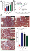
Viruses differ in their ability to exploit GFP-LC3, and there appears to be a threshold effect. Two approaches were used to determine if the beneficial effects of host-derived and virus-delivered LC3 were additive. First, WT mice (black bars) or GFP-Lc3 mice (green hatched bars) carrying a single allele of GFP-LC3 were infected with the 4 indicated rCVB3s, or with WT CVB3, and were sacrificed at d 2 p.i. (A) Pancreatic viral titers were determined, and (B) for the rCVB3s, RNA genome quantities were measured using RT-PCR. Second (C), the replication of the 4 rCVB3s was evaluated in mice homozygous for GFP-Lc3 (i.e, carrying 2 alleles of the transgene, solid green bars) and was compared to the titers in mice with a single gene copy. (D) Upper panel: Mice hemizygous (WT/tg) and homozygous (tg/tg) for GFP-Lc3 were genotyped as described. To determine if the GFP-Lc3 gene copy number was reflected in the amount of GFP-LC3 protein that was present, western blots were carried out on pancreatic protein extracts, and probed using an antibody to LC3, or to GAPDH (loading control). After normalizing for the amounts of GAPDH, the amount of GFP-LC3 in pancreata of tg/tg mice was found to be ∼2.8x higher than that of their WT/tg counterparts.
Figure 6.
Figure 6.
rCVB3-encoded ATG5 restores the functional deficiency in ElCre-Atg5f/f acinar cells, and enhances viral replication and pathogenesis. (A) The extent, and cell-specificity, of ELCre activity was determined by confocal microscopy of vibratome sections from the F1 cross of ElCre-Atg5f/f mice against JAX 7914 mice (which carry a reporter cassette that, when cleaved by Cre recombinase, causes expression of the Tomato fluorescent protein). Genotyping showed that, as expected, all mice in the resulting litter were Atg5f/WT-7914+; and ∼50% also carried ElCre. Pancreatic samples were taken from mice expressing ELCre (2 central images, the rightmost of which shows a higher magnification including an islet of Langerhans). Negative controls were (i) pancreatic tissue from a mouse lacking ElCre (left image) and (ii) liver from an ELCre-expressing mouse. Red = Tomato; Blue = nuclei (Hoechst 33342 dye); Green = F-actin (phalloidin staining; phalloidin binds to F-actin, thereby revealing the cytoskeleton). Images are representative, and are from 2 independent experiments (n = 4 mice per group). Twenty-six pancreatic fields were evaluated for ElCre mice, and 2 fields for the Cre-negative control mice. (B) HeLa cells were infected with Atg5-CVB3 or with dsRed-CVB3, and a western blot was probed for ATG5, or for GAPDH. (C) ElCre-Atg5f/f mice were infected with Atg5-CVB3 or with dsRed-CVB3 and, 2 d later, their pancreata were harvested, and the amount of ATG12-ATG5 complex was determined by western blot. The complex is absent from the pancreata of mice infected with dsRed-CVB3, but is abundant in the pancreata of Atg5-CVB3-infected animals, demonstrating that the virus-encoded ATG5 is biologically active. (D). Virus titers show that the provision of ATG5 by rCVB3 increases viral replication in the pancreata of ElCre-Atg5f/f mice. (E). This increased replication is reflected by marked pancreatitis (Masson trichrome stain; n = 7 mice, in 5 independent experiments; quantitation shown in bar graph).
Figure 7.
Figure 7.
rCVB3s encoding different autophagy-related proteins show differing dependence on an intact autophagy pathway: evaluating the involvement of SEL1L. (A) To determine if the beneficial impact of autophagy-related proteins was dependent on an intact autophagy pathway, ElCre-Atg5f/f mice were infected with each of the 3 new rCVB3s, or with dsRed-CVB3 as a control. Pancreata were harvested at d 1 or 2 p.i., and virus titers were determined. Next, for reasons explained in the text, we investigated the binding of LC3-related proteins to the ERAD pathway protein SEL1L. HeLa cell lysates were prepared from uninfected cells (Un) and from cells infected by each of the 4 indicated rCVB3. (B) Aliquots of these lysates were subjected to gel electrophoresis, and western blots were carried out using antibody to LC3, or to GAPDH (loading control). As was explained for Figure 2, the great abundance of LC3-related proteins produced by the proLC3 and proLC3G120A viruses required that less total protein be applied to those lanes. Identification of each band is facilitated by the inclusion of a colored dot; the data are nearly identical to those shown in Figure 2D. (C) Equal amounts of each protein lysate were immunoprecipitated with antibody to SEL1L. The precipitates were subjected to gel electrophoresis, and western blots were carried out using antibody to LC3, or to SEL1L. The observed coimmunoprecipitation of LC3 and SEL1L was confirmed in 5 independent experiments. (D) As an additional control for the specificity of immunoprecipitation (see text), the lysates were immunoprecipitated using an antibody to the ER resident protein CANX then, after electrophoresis and blotting, were probed for the presence of LC3-related proteins (top panel) or CANX (bottom panel).
Figure 8.
Figure 8.
Diagrammatic summary: CVB3 can exploit cellular membranes from a variety of sources. We propose that CVB3 can exploit 3 distinct sources of membrane scaffolds, all arising from the ER. Two of them are constitutive cellular pathways (autophagy and ERAD), and both of them (i) involve the attachment, covalently or otherwise, of LC3 to the ER membrane; and (ii) culminate in the production of abundant DMVs. The third pathway is extant only when LC3-I is sequestered by ATG4BC74A, giving rise to immature phagophores and reorganized membranes. Only the first of these 3 pathways is ATG5-dependent.
None

References

    1. Uetz P, Dong YA, Zeretzke C, Atzler C, Baiker A, Berger B, Rajagopala SV, Roupelieva M, Rose D, Fossum E, et al.. Herpesviral protein networks and their interaction with the human proteome. Science 2006; 311:239-42; PMID:16339411; http://dx.doi.org/10.1126/science.1116804 - DOI - PubMed
    1. Miller S, Krijnse-Locker J. Modification of intracellular membrane structures for virus replication. Nat Rev Microbiol 2008; 6:363-74; PMID:18414501; http://dx.doi.org/10.1038/nrmicro1890 - DOI - PMC - PubMed
    1. Kirkegaard K. Subversion of the cellular autophagy pathway by viruses. Curr Top Microbiol Immunol 2009; 335:323-33; PMID:19802573 - PubMed
    1. Dales S, Eggers HJ, Tamm I, Palade GE. Electron microscopic study of the formation of poliovirus. Virology 1965; 26:379-89; PMID:14319710; http://dx.doi.org/10.1016/0042-6822(65)90001-2 - DOI - PubMed
    1. Jezequel AM, Steiner JW. Some ultrastructural and histochemical aspects of Coxsackie virus-cell interactions. Lab Invest 1966; 15:1055-83; PMID:5911943 - PubMed

Publication types

MeSH terms

LinkOut - more resources