. 2015 Jun 19;10(6):e0131062.
doi: 10.1371/journal.pone.0131062. eCollection 2015.

Constitutive Activity in an Ancestral Form of Abl Tyrosine Kinase

Affiliations

Constitutive Activity in an Ancestral Form of Abl Tyrosine Kinase

Saadat U Aleem et al. PLoS One. .

Abstract

The c-abl proto-oncogene encodes a nonreceptor tyrosine kinase that is found in all metazoans, and is ubiquitously expressed in mammalian tissues. The Abl tyrosine kinase plays important roles in the regulation of mammalian cell physiology. Abl-like kinases have been identified in the genomes of unicellular choanoflagellates, the closest relatives to the Metazoa, and in related unicellular organisms. Here, we have carried out the first characterization of a premetazoan Abl kinase, MbAbl2, from the choanoflagellate Monosiga brevicollis. The enzyme possesses SH3, SH2, and kinase domains in a similar arrangement to its mammalian counterparts, and is an active tyrosine kinase. MbAbl2 lacks the N-terminal myristoylation and cap sequences that are critical regulators of mammalian Abl kinase activity, and we show that MbAbl2 is constitutively active. When expressed in mammalian cells, MbAbl2 strongly phosphorylates cellular proteins on tyrosine, and transforms cells much more potently than mammalian Abl kinase. Thus, MbAbl2 appears to lack the autoinhibitory mechanism that tightly constrains the activity of mammalian Abl kinases, suggesting that this regulatory apparatus arose more recently in metazoan evolution.


Conflict of interest statement

Competing Interests: The authors have declared that no competing interests exist.

Figures

Fig 1
Fig 1. Domain organization of MbAbl2.
(A) A schematic diagram showing the domain organization of MbAbl2 and mammalian Abl (murine c-Abl, 1b isoform). The percent amino acid identity for each domain is shown below the figure. (B) Cartoon representing the autoinhibited structure of mammalian Abl. The SH3 domain is in yellow, the SH2 domain is green, and the kinase domain is blue. The N-terminal myristoyl group (Myr) binds in a pocket on the kinase domain. The cap sequence is shown by a dashed red line, and the intramolecular SH3 ligand is shown by a solid red line. Tyrosine 412 (Y) in the activation loop is pictured. (C) Sequence comparison at the autophosphorylation site within the kinase activation loop.
Fig 2
Fig 2. Enzymatic activity of MbAbl2.
(A) SDS-PAGE analysis of purified MbAbl2, detected by Coomassie blue staining (left) or by anti-His-tag Western blotting (right). (B) Initial rates of substrate phosphorylation for various concentrations of MbAbl2 were measured in triplicate by the continuous spectrophotometric assay. The peptide substrate used, Abl peptide (EAIYAAPFAKKKG), was at a concentration of 100 μM. Error bars show standard deviations. (C) MbAbl2 initial rates were measured at varying concentrations of ATP using the continuous spectrophotometric assay. The fit to a hyperbolic substrate vs. velocity curve is shown. (D) MbAbl2-catalyzed phosphorylation of peptide substrates containing recognition motifs for various kinases. The peptide sequences are given in the Materials and Methods section. Enzymatic activity was measured using the phosphocellulose paper binding assay, with an enzyme concentration of 275 nM and peptide concentrations of 500 μM. Reactions proceeded for 10 minutes at 30°C. Error bars show standard deviation.
Fig 3
Fig 3. Effect of kinase inhibitors on MbAbl2.
MbAbl2 activity was measured in the presence of 1 μM and 20 μM concentrations of the indicated inhibitors. Activity was measured using the phosphocellulose binding assay with Abl peptide substrate at a concentration of 125 μM. The concentration of ATP was 200 μM. Error bars show standard deviations.
Fig 4
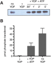
Fig 4. Regulation of MbAbl2 by autophosphorylation.
(A) Purified MbAbl2 (200 μl of 825 nM) was incubated with immobilized GST-YOP tyrosine phosphatase (200 μl) for 45 minutes at room temperature, or left untreated (no YOP). Following removal of GST-YOP by centrifugation, autophosphorylation was initiated by the addition of ATP (0.5 mM) and MgCl2 (20 mM). After various reaction times (at room temperature), aliquots were removed and immediately quenched by addition of Laemmli sample buffer and boiling. The samples were analyzed by SDS-PAGE and anti-pTyr Western blotting; equal amounts of MbAbl2 (14 pmol) were analyzed in each lane. (B) YOP treatment and autophosphorylation were carried out as in panel (A). After 30 min. of autophosphorylation, MbAbl2 activity was measured using the phosphocellulose paper binding assay with Abl peptide (740 μM). Kinase reactions proceeded for 10 minutes at 30°C. Error bars show standard deviations.
Fig 5
Fig 5. Abl activity in mammalian cells.
(A) Lysates from NIH3T3 cells stably expressing Abl, MbAbl2, or empty vector (EV) (100 μg) were separated by SDS-PAGE and analyzed by anti-pTyr Western blotting. The membrane was stripped and reprobed with anti-Flag and tubulin antibodies to ensure equal loading of Abl and MbAbl2. The figure is representative of three independent experiments. (B) Lysates from NIH3T3 cells expressing Abl-Flag or MbAbl2-Flag (1 mg total protein) were subjected to immunoprecipitation reactions with anti-Flag affinity resin. Duplicate samples of the precipitated proteins were used in an in vitro kinase reaction with Abl peptide and [γ -32P]-ATP. Error bars show standard deviations. A separate sample was analyzed by Western blotting with anti-Flag antibody (inset). (C) Colony formation assay. NIH3T3 cells expressing Abl, MbAbl2, or vector control were sorted for equal GFP expression. Cells (1000 per well) were resuspended in low melting point agarose prepared in complete media. Wells were photographed after 28 days. This experiment was repeated three times with similar results. (D) Anchorage-independent growth assay. NIH3T3 cells expressing Abl, MbAbl2, or vector control (20,000 per well; dashed line) were resuspended in complete media and added to ultra low attachment plates. Cells were counted after 6 days of growth. This experiment was performed three times with similar results. Error bars show standard deviations.

References

    1. Colicelli J ABL tyrosine kinases: evolution of function, regulation, and specificity. Sci Signal 3: re6 10.1126/scisignal.3139re6 - DOI - PMC - PubMed
    1. Nagar B, Hantschel O, Young MA, Scheffzek K, Veach D, Bornmann W, et al. (2003) Structural basis for the autoinhibition of c-Abl tyrosine kinase. Cell 112: 859–871. - PubMed
    1. Wong S, Witte ON (2004) The BCR-ABL story: bench to bedside and back. Annu Rev Immunol 22: 247–306. - PubMed
    1. Druker BJ, Lydon NB (2000) Lessons learned from the development of an abl tyrosine kinase inhibitor for chronic myelogenous leukemia. J Clin Invest 105: 3–7. - PMC - PubMed
    1. Daley GQ, Van Etten RA, Baltimore D (1990) Induction of chronic myelogenous leukemia in mice by the P210bcr/abl gene of the Philadelphia chromosome. Science 247: 824–830. - PubMed

Publication types

MeSH terms

LinkOut - more resources