Case Reports
. 2016 Jan;54(1):71-80.
doi: 10.1165/rcmb.2015-0154OC.

Deficiency Mutations of Alpha-1 Antitrypsin. Effects on Folding, Function, and Polymerization

Affiliations
Case Reports

Deficiency Mutations of Alpha-1 Antitrypsin. Effects on Folding, Function, and Polymerization

Imran Haq et al. Am J Respir Cell Mol Biol. 2016 Jan.

Abstract

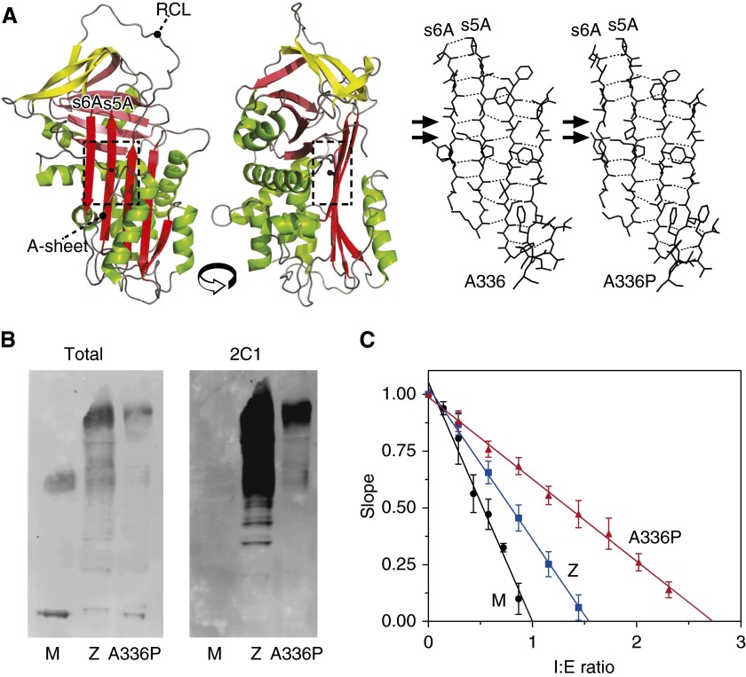
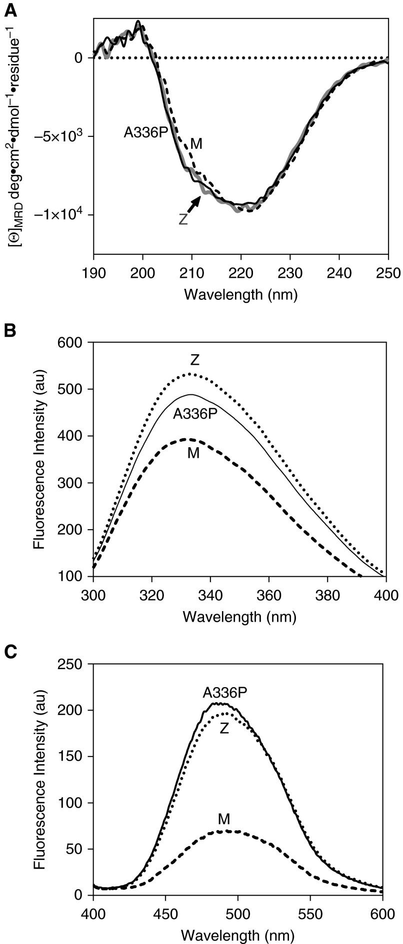
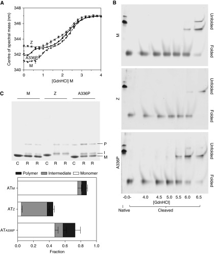
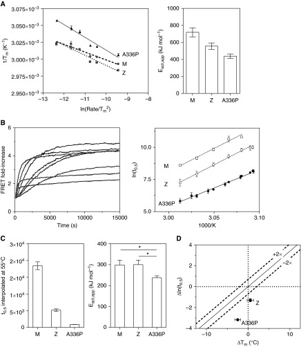
Misfolding, polymerization, and defective secretion of functional alpha-1 antitrypsin underlies the predisposition to severe liver and lung disease in alpha-1 antitrypsin deficiency. We have identified a novel (Ala336Pro, Baghdad) deficiency variant and characterized it relative to the wild-type (M) and Glu342Lys (Z) alleles. The index case is a homozygous individual of consanguineous parentage, with levels of circulating alpha-1 antitrypsin in the moderate deficiency range, but is a biochemical phenotype that could not be classified by standard methods. The majority of the protein was present as functionally inactive polymer, and the remaining monomer was 37% active relative to the wild-type protein. These factors combined indicate an 85 to 95% functional deficiency, similar to that seen with ZZ homozygotes. Biochemical, biophysical, and computational studies further defined the molecular basis of this deficiency. These studies demonstrated that native Ala336Pro alpha-1 antitrypsin could populate the polymerogenic intermediate-and therefore polymerize-more readily than either wild-type alpha-1 antitrypsin or the Z variant. In contrast, folding was far less impaired in Ala336Pro alpha-1 antitrypsin than in the Z variant. The data are consistent with a disparate contribution by the "breach" region and "shutter" region of strand 5A to folding and polymerization mechanisms. Moreover, the findings demonstrate that, in these variants, folding efficiency does not correlate directly with the tendency to polymerize in vitro or in vivo. They therefore differentiate generalized misfolding from polymerization tendencies in missense variants of alpha-1 antitrypsin. Clinically, they further support the need to quantify loss-of-function in alpha-1 antitrypsin deficiency to individualize patient care.

Keywords: alpha-1 antitrypsin deficiency; mutation; polymerization; serpinopathies; unfolding.


Figures

Figure 1.
Figure 1.
Characteristics of patient-derived alpha-1 antitrypsin. (A) Left panels: Structure cartoon of the wild-type protein (accession 1QLP [18]) generated using PyMol (46) showing the position of Ala336 in the standard β-sheet A (A-sheet) view and rotated counterclockwise by 90°, with the reactive center loop (RCL) indicated. Sheets A, B, and C are colored red, pink, and yellow, respectively. Right panels: An energy-minimized molecular model of ATA336P compared with the wild-type coordinates treated in the same manner using NAMD (20) and VMD (19). The arrows indicate the loss of two main-chain hydrogen bonds (hatched lines) in the model between strands 5A and 6A. (B) Western blot analysis of patient-derived plasma from MM, ZZ, and Ala336Pro/Ala336Pro homozygotes separated by 3 to 12% wt/vol nondenaturing PAGE with detection by rabbit polyclonal (Total) and polymer-specific (2C1) antibodies. Higher-order bands represent polymeric species. The ATA336P polymer is recognized by 2C1, demonstrating the presence of an epitope shared with the Z polymer. (C) Calculation of the stoichiometries of inhibition of alpha-1 antitrypsin variants against bovine α-chymotrypsin showing residual activity at different ratios of inhibitor to enzyme (I:E ratio). Error bars denote SDs from three experiments.
Figure 2.
Figure 2.
Structural features of Ala336Pro. (A) Assessment of overall secondary structure by far-ultraviolet circular dichroism (CD). CD spectra of plasma-derived alpha-1 antitrypsin, at 0.5 mg/ml in 10 mM Na2HPO4/NaH2PO4 (pH 7.4) recorded between 250 and 190 nm show similar profiles for the three variants. (B) Assessment of breach opening (30) by intrinsic tryptophan fluorescence. Spectra were recorded of 0.5 μM alpha-1 antitrypsin in PBS with 5% vol/vol glycerol between 300 and 400 nm with excitation at 295 nm. Ala336Pro shows increased fluorescence intensity and red shift in wavelength compared with ATM but to a lesser extent than ATZ alpha-1 antitrypsin. (C) Alpha-1 antitrypsin (2 μM) in PBS with 5% vol/vol glycerol was incubated with 10 μM 4,4′-dianilino-1,1′-binaphthyl-5,5′-disulfonic acid for 10 minutes, and the spectra were recorded between 400 and 600 nm with excitation at 370 nm. The increase in fluorescence was comparable to ATZ, suggesting increased population of the polymerization intermediate (34). [Θ]MRD, mean residue ellipticity.
Figure 3.
Figure 3.
Equilibrium unfolding and rapid refolding of alpha-1 antitrypsin. (A) Changes in intrinsic fluorescence center of spectral mass during equilibrium unfolding of 0.5 μM alpha-1 antitrypsin are shown. Values calculated at 6 M guanidinium hydrochloride (GdnHCl) were similar to those at 4 M. Samples were in 10 mM Na2HPO4/NaH2PO4 (pH 7.4). (B) ATM, ATZ, and ATA336P were cleaved overnight at a 50-fold molar excess with respect to Staphylococcus aureus protease V8 and incubated for 2 hours in increasing concentrations of guanidinium hydrochloride before separation by 6 M urea PAGE. In each 8% acrylamide gel, native and cleaved proteins in the absence of denaturant (left two lanes) were used as controls. Unfolding of the protein resulted in a decreased rate of migration. This shift in behavior occurred at a lower concentration for ATA336P than the other variants. (C) Top: alpha-1 antitrypsin (1 μg) was concentrated to 10 mg/ml in PBS with 5% vol/vol glycerol (C) or in 6 M guanidinium hydrochloride (R) before 200-fold dilution into PBS with 5% vol/vol glycerol. Samples were resolved on a 3 to 12% wt/vol nondenaturing gel stained with Coomassie Brilliant Blue. M, I, and P represent the positions of monomer, intermediate, and polymer fractions, respectively (42). Bottom: Analysis of refolding by densitometry. Each bar represents the total protein regained on refolding compared with the control (C) samples. Demarcation of the bars represents the proportion of conformer regained.
Figure 4.
Figure 4.
The effects of Ala336Pro on thermal stability. (A) Left: Conversion of ATM, ATZ, and ATA336P variants from native to intermediate upon heating between 25 and 95°C was monitored in PBS using SYPRO Orange dye (21), and the midpoint of the thermal transition (Tm) was plotted as a function of rate of temperature increase (47). Right: The calculated apparent activation energy values (Eact,app) for the transition between native and intermediate states, calculated from the slopes of the regressions. (B) Left: ATA336P, labeled with Alexa 488 and Alexa 594 dyes at Cys232 in equimolar ratios, was heated over a range of temperatures at 0.1 mg/ml in PBS, and the increase in FRET (Förster Resonance Energy Transfer) was monitored. Typical results of an experiment are shown (solid black lines), with curves of best fit (dashed gray lines). Right: Half-times of polymerization of ATA336P were determined from FRET progress curves at a range of temperatures and subjected to an Årrhenius analysis (17, 21). (C) Left: The Årrhenius plot was used to interpolate a polymerization half-time for the variants at a single reference temperature (55°C). The polymerization of ATA336P was faster than for ATZ or ATM variants. Right: The apparent activation energy for polymerization, Eact,app, was calculated from the Årrhenius plot. This was significantly lower for ATA336P than for the ATM or ATZ variants as judged by a one-way ANOVA (P < 0.05). (D) Half-times of polymerization at 55°C and the Tm values of the ATZ and ATA336P variants are shown relative to that of ATM. The dashed lines represent a consensus relationship between a rate of polymerization dictated by native state stability (21).

References

    1. Zaimidou S, van Baal S, Smith TD, Mitropoulos K, Ljujic M, Radojkovic D, Cotton RG, Patrinos GP. A1ATVar: a relational database of human SERPINA1 gene variants leading to α1-antitrypsin deficiency and application of the VariVis software. Hum Mutat. 2009;30:308–313. - PubMed
    1. Brantly ML, Wittes JT, Vogelmeier CF, Hubbard RC, Fells GA, Crystal RG. Use of a highly purified alpha 1-antitrypsin standard to establish ranges for the common normal and deficient alpha 1-antitrypsin phenotypes. Chest. 1991;100:703–708. - PubMed
    1. Allan PC, Harley RA, Talamo RC. A new method for determination of alpha-1-antitrypsin phenotype using isoelectric focusing on polyacrylamide gel slabs. Am J Clin Pathol. 1974;62:732–739. - PubMed
    1. Sveger T. Liver disease in alpha1-antitrypsin deficiency detected by screening of 200,000 infants. N Engl J Med. 1976;294:1316–1321. - PubMed
    1. Silverman EK, Miletich JP, Pierce JA, Sherman LA, Endicott SK, Broze GJ, Jr, Campbell EJ. Alpha-1-antitrypsin deficiency: high prevalence in the St. Louis area determined by direct population screening. Am Rev Respir Dis. 1989;140:961–966. - PubMed

Publication types

MeSH terms

Substances