. 2015 Aug 3;14(15):2473-83.
doi: 10.1080/15384101.2015.1005530. Epub 2015 Jun 19.

Exosomes derived from human mesenchymal stem cells confer drug resistance in gastric cancer

Affiliations

Exosomes derived from human mesenchymal stem cells confer drug resistance in gastric cancer

Runbi Ji et al. Cell Cycle. .

Abstract

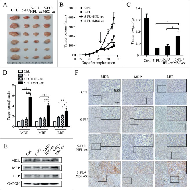
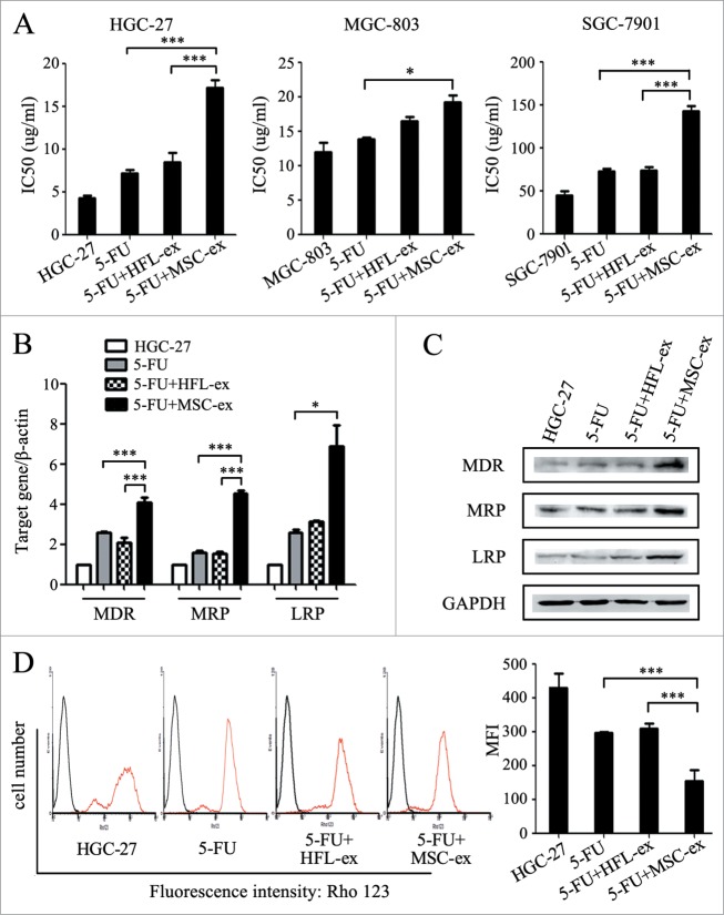
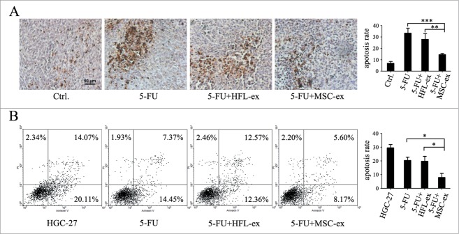
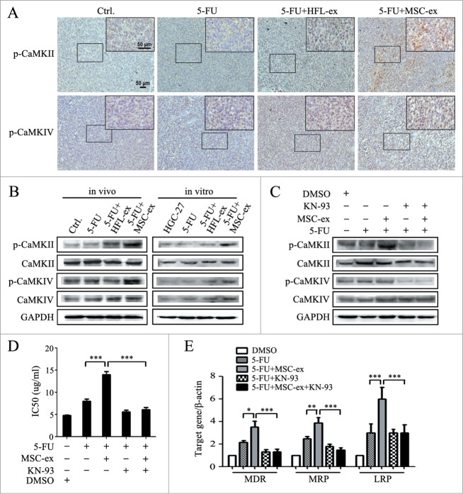
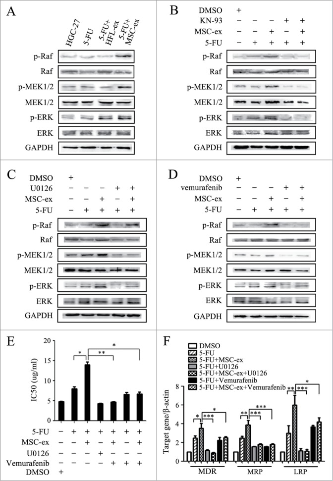
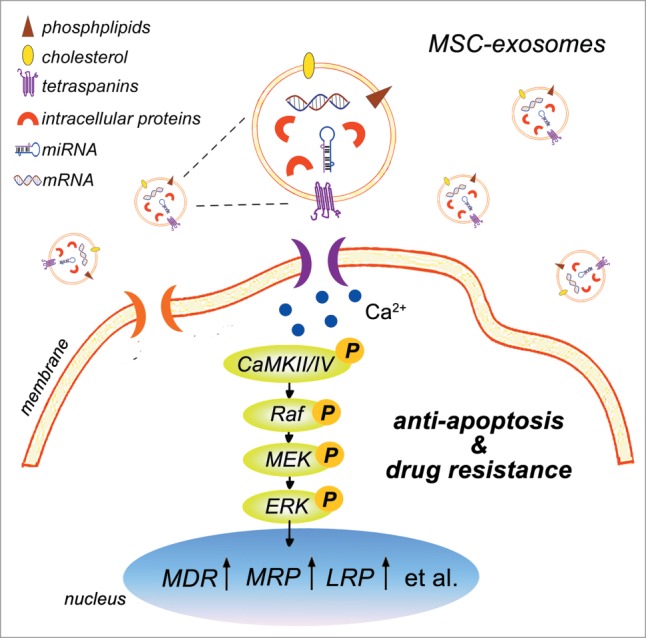
Mesenchymal stem cells (MSCs) play an important role in chemoresistance. Exosomes have been reported to modify cellular phenotype and function by mediating cell-cell communication. In this study, we aimed to investigate whether exosomes derived from MSCs (MSC-exosomes) are involved in mediating the resistance to chemotherapy in gastric cancer and to explore the underlying molecular mechanism. We found that MSC-exosomes significantly induced the resistance of gastric cancer cells to 5-fluorouracil both in vivo and ex vivo. MSC-exosomes antagonized 5-fluorouracil-induced apoptosis and enhanced the expression of multi-drug resistance associated proteins, including MDR, MRP and LRP. Mechanistically, MSC-exosomes triggered the activation of calcium/calmodulin-dependent protein kinases (CaM-Ks) and Raf/MEK/ERK kinase cascade in gastric cancer cells. Blocking the CaM-Ks/Raf/MEK/ERK pathway inhibited the promoting role of MSC-exosomes in chemoresistance. Collectively, MSC-exosomes could induce drug resistance in gastric cancer cells by activating CaM-Ks/Raf/MEK/ERK pathway. Our findings suggest that MSC-exosomes have profound effects on modifying gastric cancer cells in the development of drug resistance. Targeting the interaction between MSC-exosomes and cancer cells may help improve the efficacy of chemotherapy in gastric cancer.

Keywords: drug resistance; exosomes; gastric cancer; mesenchymal stem cells.


Figures

Figure 1.
Figure 1.
MSC-exosomes induce resistance of gastric cancer cells to 5-FU in vivo. (A) The size of tumors at the end of the experiment from mice treated with PBS (Ctrl.), 5-FU, 5-FU+HFL1-exosomes, 5-FU+MSC-exosomes. (B) Tumor growth curves in mice treated with PBS, 5-FU, 5-FU+HFL1-exosomes, or 5-FU+MSC-exosomes (n=6). Treatment was initiated when tumors reached a volume of 50–100 mm3. (C) The mean weight of tumors at the end of the experiment (at day 7 after chemotherapy treatment) from mice treated with PBS, 5-FU, 5-FU+HFL1-exosomes, or 5-FU+MSC-exosomes (n=6). (* P<0.05). (D) Relative quantitative PCR analyses of MDR, MRP, and LRP gene expression in tumor tissues from mice treated with PBS, 5-FU, 5-FU+HFL1-exosomes, or 5-FU+MSC-exosomes. (* P<0.05, ** P<0.01, *** P<0.001.) (E) The expression levels of MDR, MRP, and LRP proteins were examined by using western blot. (F) Immunohistochemical analyses of MDR, MRP and LRP protein expression in tumor tissues from mice treated with PBS, 5-FU, 5-FU+HFL1-exosomes, or 5-FU+MSC-exosomes. Original magnification, × 100, smaller one at top right corner, × 200. Scale bar = 50 μm.
Figure 2.
Figure 2.
MSC-exosomes induce resistance of gastric cancer cells to 5-FU ex vivo. (A) MTT assay for IC50 of parental and chemoresistant HGC-27, MGC-803, and SGC-7901 cells in response to 5-FU. The cells were treated with 5-FU for 24 h, then changed to normal medium until cell recovery. Exosomes from MSCs and HFL1 cells were added at the start of treatment for 72 h. The control cells were cultured in normal medium without any treatment. (* P < 0.05, *** P < 0.001). (B) The expression of MDR, MRP, and LRP genes in parental and chemoresistant HGC-27 cells was determined by using relative quantitative PCR. (* P < 0.05, *** P < 0.001). (C) Western blot assays for MDR, MRP and LRP protein expression in parental and chemoresistant HGC-27 cells. (D) Fluorescent intensity of Rho-123 in parental and chemoresistant HGC-27 cells. The cells were labeled with Rho-123 after exposure to 5-FU for 6 h (red line). The cells without Rho-123 labeling were used as control (black line). For each assay, 10,000 cells were analyzed. The x-axis corresponds to the fluorescence intensity, and the y-axis, to the number of cells per channel. The quantitative data are presented as the mean ± SD of triplicate experiments. MFI: the mean fluorescent intensity. (*** P < 0.001).
Figure 3.
Figure 3.
MSC-exosomes protect gastric cancer cells from chemotherapy-induced apoptosis. (A) Tumors from mice treated with PBS (Ctrl.), 5-FU, 5-FU+HFL1-exosomes, 5-FU+MSC-exosomes were paraffin-embedded and sectioned, followed by staining of apoptotic cell by using TUNEL assay. The number of TUNEL-positive cells notably increased in the 5-FU and 5-FU+HFL1-exosome groups compared to the 5-FU+MSC-exosome group, while the control group treated with PBS had few apoptotic cells. Original magnification, × 200. Scale bar = 50 μm. The quantitative analyses of apoptosis (TUNEL) indices were calculated by counting the number of positive cells in 10 random fields. (** P < 0.01, *** P < 0.001). (B) Flow cytometric analyses of apoptotic cells ex vivo. The parental and chemoresistant HGC-27 cells were exposed to 5-FU for 48 h, collected and subjected to Annexin V/PI double staining, followed by FACS analyses. For each assay, 10,000 cells were analyzed. The quantitative data are presented as the mean ± SD of triplicate experiments. (* P < 0.05).
Figure 4.
Figure 4.
MSC-exosomes activate CaM-KII and CaM-KIV in gastric cancer cells. (A) Tumors from mice treated with PBS (Ctrl.), 5-FU, 5-FU+HFL1-exosomes, 5-FU+MSC-exosomes were paraffin-embedded and sectioned, followed by immunohistochemical staining of p-CaM-KII and p-CaM-KIV. Original magnification, × 100, smaller one at top right corner, × 200. Scale bar = 50 μm. (B) Western blot analyses of CaM-KII, CaM-KIV, and their phosphorylated forms in tumor tissues and cells. (C) HGC-27 cells were treated with MSC-exosomes in the presence or absence of KN-93 (10 μM). The protein levels of p-CaM-KII and p-CaM-KIV were examined by using protein gel blot. (D, E) HGC-27 cells were treated with MSC-exosomes in the presence or absence of KN-93 (10 μM). The IC50 of HGC-27 cells in response to 5-FU were determined by using MTT assay (D). The expression of MDR, MRP, and LRP genes was determined by using relative quantitative PCR (E). (* P < 0.05, ** P < 0.01, *** P < 0.001).
Figure 5.
Figure 5.
Activation of the CaM-Ks/Raf/MEK/ERK pathway is critical for chemoresistance induced by MSC-exosomes. (A) The expression of p-Raf, p-MEK, and p-ERK in the parental and chemoresistant HGC-27 cells was detected by using western blot. (B) Western blot assays for the expression of p-Raf, p-MEK, and p-ERK in HGC-27 cells treated with MSC-exosomes in the presence or absence of KN-93 (10 μM). (C) HGC-27 cells were treated with MSC-exosomes in the presence or absence of U0126 (10 μM). The levels of phosphorylated Raf, MEK, and ERK were examined by using protein gel blot. (D) HGC-27 cells were treated with MSC-exosomes together with or without vemurafenib (20 μM). The expression of p-Raf, p-MEK, and p-ERK was examined by using western blot. (E, F) HGC-27 cells were treated with MSC-exosomes in the presence or absence of U0126 (10 μM) or vemurafenib (20 μM). The IC50 of HGC-27 cells in response to 5-FU was determined by using MTT assay (E). The expression of MDR, MRP, and LRP genes was determined by using relative quantitative PCR (F). (* P < 0.05, ** P < 0.01, *** P < 0.001).
Figure 6.
Figure 6.
MSC-exosomes induce chemoresistance in gastric cells mainly though proteins. (A) MTT assay for IC50 of HGC-27 cells in response to 5-FU. MSC-exosomes were pre-incubated with nucleic acid-hydrolyzing enzyme (RNase A) and proteolytic enzyme (Proteinase K). HGC-27 were treated with 5-FU for 24 h, then changed to normal medium until cell recovery. The pre-treated and untreated MSC-exosomes were added at the start of 5-FU treatment for 72 h. The control cells were cultured in normal medium without any treatment. (** P < 0.01, *** P < 0.001). (B) The expression of MDR, MRP, and LRP genes in HGC-27 was determined by using relative quantitative PCR. (* P < 0.05, ** P < 0.01, *** P < 0.001). (C) Western blot assays for MDR, MRP and LRP proteins expression in HGC-27 cells. (D) MSCs were transfected with negative control (NC) and siRNA-MDR by using Lipofectin. The expression of MDR in MSCs and MSC-exosomes were determined by using protein gel blot. (E) MTT assay for IC50 of parental and chemoresistant HGC-27 cells to 5-FU. (* P < 0.05, ** P < 0.01). (F) Relative quantitative PCR analyses of MDR, MRP, and LRP genes expression. (** P<0.01, *** P<0.001). (G) The expression levels of MDR, MRP, and LRP proteins were examined by using western blot.
Figure 7.
Figure 7.
Schematic model for the role of MSC-exosomes in the development of drug resistance in gastric cancer. MSC-exosomes contains a variety of bioactive molecules ranging from mRNAs, proteins, to miRNAs. MSC-exosomes incorporated into the gastric cancer cells stimulate the activation of CaM-Ks (predominantly CaM-KII and CaM-KIV). Activation of CaM-KII and CaM-KIV trigger the activation of downstream Raf/MEK/ERK signaling cascade. Consequently, the expression of multi-drug resistant proteins is up-regulated in gastric cancer cells, resulting in the resistance to chemotherapy-induced apoptosis and the development of drug resistance.

References

    1. Roodhart JM, Daenen LG, Stigter EC, Prins HJ, Gerrits J, Houthuijzen JM, Gerritsen MG, Schipper HS, Backer MJ, van Amersfoort M, et al. . Mesenchymal stem cells induce resistance to chemotherapy through the release of platinum-induced fatty acids. Cancer Cell 2011; 20(3):370-83; PMID:21907927; http://dx.doi.org/10.1016/j.ccr.2011.08.010 - DOI - PubMed
    1. Cukierman E, Bassi DE. The mesenchymal tumor microenvironment: a drug-resistant niche. Cell Adh Migr 2012; 6(3):285-96; PMID:22568991; http://dx.doi.org/10.4161/cam.20210 - DOI - PMC - PubMed
    1. Zhu W, Huang L, Li Y, Qian H, Shan X, Yan Y, Mao F, Wu X, Xu WR. Mesenchymal stem cell-secreted soluble signaling molecules potentiate tumor growth. Cell Cycle 2011; 10(18):3198-207; PMID:21900753; http://dx.doi.org/10.4161/cc.10.18.17638 - DOI - PubMed
    1. Zhu W, Huang L, Li Y, Zhang X, Gu J, Yan Y, Xu X, Wang M, Qian H, Xu W. Exosomes derived from human bone marrow mesenchymal stem cells promote tumor growth in vivo. Cancer Lett 2012; 315(1):28-37; PMID:22055459; http://dx.doi.org/10.1016/j.canlet.2011.10.002 - DOI - PubMed
    1. Li T, Yan Y, Wang B, Qian H, Zhang X, Shen L, Wang M, Zhou Y, Zhu W, Li W, et al. . Exosomes derived from human umbilical cord mesenchymal stem cells alleviate liver fibrosis. Stem Cells Dev 2013; 22(6):845-54; PMID:23002959; http://dx.doi.org/10.1089/scd.2012.0395 - DOI - PMC - PubMed

Publication types

MeSH terms