. 2015 Jun 18;58(6):911-24.
doi: 10.1016/j.molcel.2015.06.012.

The Promise of Proteomics for the Study of ADP-Ribosylation

Affiliations

The Promise of Proteomics for the Study of ADP-Ribosylation

Casey M Daniels et al. Mol Cell. .

Abstract

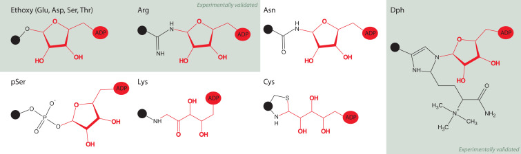
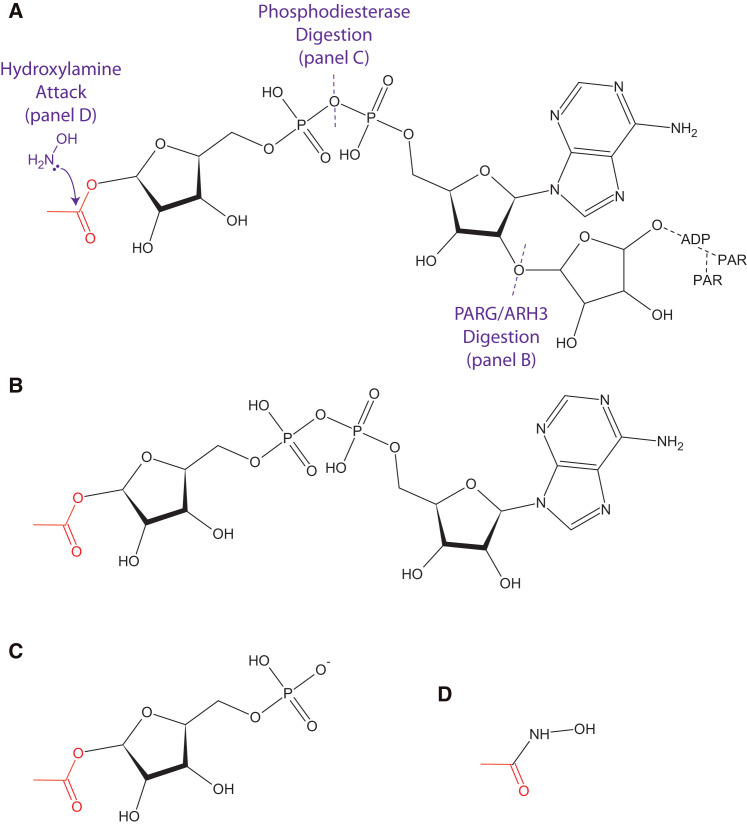
ADP-ribosylation is a post-translational modification where single units (mono-ADP-ribosylation) or polymeric chains (poly-ADP-ribosylation) of ADP-ribose are conjugated to proteins by ADP-ribosyltransferases. This post-translational modification and the ADP-ribosyltransferases (also known as PARPs) responsible for its synthesis have been found to play a role in nearly all major cellular processes, including DNA repair, transcription, translation, cell signaling, and cell death. Furthermore, dysregulation of ADP-ribosylation has been linked to diseases including cancers, diabetes, neurodegenerative disorders, and heart failure, leading to the development of therapeutic PARP inhibitors, many of which are currently in clinical trials. The study of this therapeutically important modification has recently been bolstered by the application of mass spectrometry-based proteomics, arguably the most powerful tool for the unbiased analysis of protein modifications. Unfortunately, progress has been hampered by the inherent challenges that stem from the physicochemical properties of ADP-ribose, which as a post-translational modification is highly charged, heterogeneous (linear or branched polymers, as well as monomers), labile, and found on a wide range of amino acid acceptors. In this Perspective, we discuss the progress that has been made in addressing these challenges, including the recent breakthroughs in proteomics techniques to identify ADP-ribosylation sites, and future developments to provide a proteome-wide view of the many cellular processes regulated by ADP-ribosylation.


Figures

Figure 1
Figure 1
The PARP Family PARPs have been linked to nearly all major cellular processes. Juxtaposition of protein identifiers (e.g., 1 = PARP-1) indicates the involvement of the protein in the regulation or execution of the cellular process. Enzymatic activity is indicated by the bubble color: blue = poly(ADP-ribosyl)transferase, red = mono(ADP-ribosyl)transferase, green = no transferase activity. For references, see Table S1.
Figure 2
Figure 2
Processes Enriched in the ADP-Ribosylated Interactome (A) Experimental design for the interactome studies used for this meta-analysis. PARGi, PARG inhibitor; PARPi, PARP inhibitor; PARGkd, PARG knockdown. (B) The pooled DNA-damaged induced ADP-ribosylated interactome depicted as a treemap of enriched biological processes. The most enriched biological processes (based on statistical likelihood) are shown as larger components within the map and grouped according to common cellular functions. See Figure S1 for the detailed version of this treemap. Gene ontology determined using DAVID (Huang et al., 2009), treemap constructed using REViGO (Supek et al., 2011) and R (R Development Core Team, 2011). (C) A compilation of the proteins identified in response to DNA damage can be broken out by enrichment methods (bait) or cell lysis conditions. For comparison of lysis conditions, the 10H enriched proteins were analyzed. Euler diagrams created in VennMaster (Kestler et al., 2005). Source data available in Table S2.
Figure 3
Figure 3
ADP-Ribosylation Attachment Sites Known and predicted structures linking amino acids to ADP-ribose, grayed out boxes show structures that have been validated. See text for references.
Figure 4
Figure 4
ADP-Ribosylation Tags (A) Poly(ADP-ribose) can be simplified to mono(ADP-ribose) as in (B) by the glycohydrolase activity of PARG/ARH3, (C) to phosphoribose through digestion by phosphodiesterase, or (D) to a hydroxamic acid derivative though exposure to hydroxylamine. Of note, hydroxylamine treatment on ADP-ribosylated arginine results in the formation of the hydroxyamate of ADP-ribose (Moss et al., 1983) and therefore will likely not leave the 15.01 Da signature on formerly modified arginine residues as in glutamate/aspartate residues (Zhang et al., 2013). A representative acidic attachment site (red) is used for illustration.
Figure 5
Figure 5
PARP Substrate Specificity Substrates for PARP-1, PARP-2, PARP-10, and PARP-14 were identified in three studies using protein arrays or analog-sensitive mutant protein identification (see text). Euler diagrams created in VennMaster. Source data available in Table S3.
Figure 6
Figure 6
PARP-1 Auto-modification Sites Schematic of PARP-1 includes protein domains and secondary structure; α helices are shown in red, β sheets in yellow. Auto-modification sites identified by at least two independent studies are shown. Size of annotated residues is based on the number of studies that have identified the modification sites. E488 and E491 located at the C terminus of the BRCT domain are identified by all MS studies and are shown as the two major auto-modification sites. Source data available in Table S4.

References

    1. Adamietz P., Hilz H. Poly(adenosine diphosphate ribose) is covalently linked to nuclear proteins by two types of bonds. Hoppe Seylers Z. Physiol. Chem. 1976;357:527–534. - PubMed
    1. Ahel I., Ahel D., Matsusaka T., Clark A.J., Pines J., Boulton S.J., West S.C. Poly(ADP-ribose)-binding zinc finger motifs in DNA repair/checkpoint proteins. Nature. 2008;451:81–85. - PubMed
    1. Ahel D., Horejsí Z., Wiechens N., Polo S.E., Garcia-Wilson E., Ahel I., Flynn H., Skehel M., West S.C., Jackson S.P. Poly(ADP-ribose)-dependent regulation of DNA repair by the chromatin remodeling enzyme ALC1. Science. 2009;325:1240–1243. - PMC - PubMed
    1. Altmeyer M., Messner S., Hassa P.O., Fey M., Hottiger M.O. Molecular mechanism of poly(ADP-ribosyl)ation by PARP1 and identification of lysine residues as ADP-ribose acceptor sites. Nucleic Acids Res. 2009;37:3723–3738. - PMC - PubMed
    1. Alvarez-Gonzalez R., Jacobson M.K. Characterization of polymers of adenosine diphosphate ribose generated in vitro and in vivo. Biochemistry. 1987;26:3218–3224. - PubMed

Publication types

MeSH terms

Substances