. 2015 Oct;21(5):544-58.
doi: 10.1007/s13365-015-0358-7. Epub 2015 Jun 20.

HIV-infected microglia mediate cathepsin B-induced neurotoxicity

Affiliations

HIV-infected microglia mediate cathepsin B-induced neurotoxicity

Frances Zenón et al. J Neurovirol. 2015 Oct.

Abstract

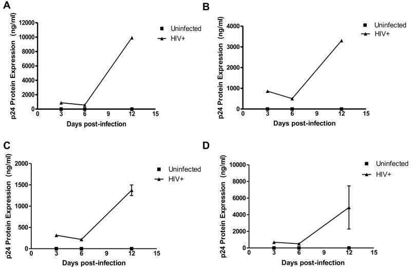
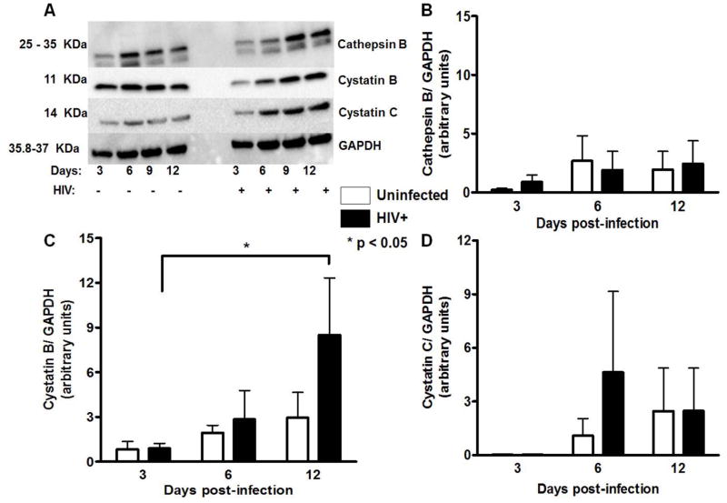
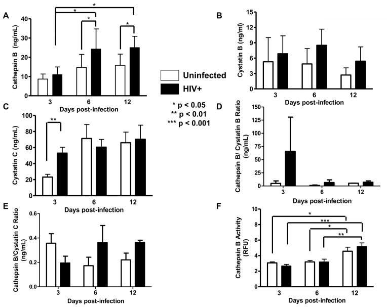
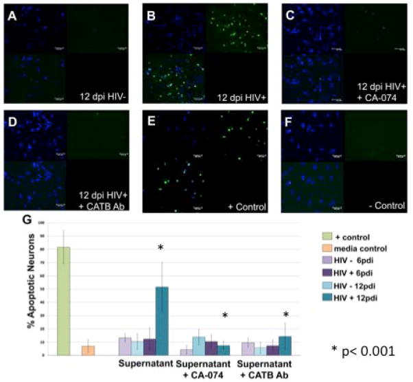
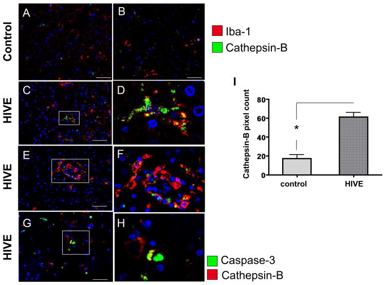
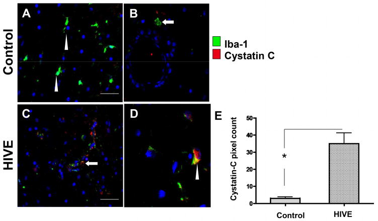
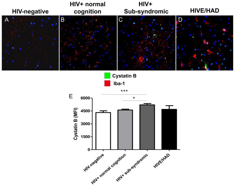
HIV-1-infected mononuclear phagocytes release soluble factors that affect the homeostasis in tissue. HIV-1 can prompt metabolic encephalopathy with the addition of neuronal dysfunction and apoptosis. Recently, we reported that HIV-1 enhances the expression and secretion of bioactive cathepsin B in monocyte-derived macrophages, ultimately contributing to neuronal apoptosis. In this research, we asked if microglia respond to HIV infection similarly by modifying the expression, secretion, and neurotoxic potential of cathepsin B and determined the in vivo relevance of these findings. HIV-1ADA-infected human primary microglia and CHME-5 microglia cell line were assessed for expression and activity of cathepsin B, its inhibitors, cystatins B and C, and the neurotoxicity associated with these changes. Human primary neurons were exposed to supernatants from HIV-infected and uninfected microglia in the presence of cathepsin B inhibitors and apoptosis was assessed by TUNEL. Microglial expression of cathepsin B was validated in brain tissue from HIV encephalitis (HIVE) patients. HIV-infected microglia secreted significantly greater levels of cathepsin B, cystatin B, and cystatin C compared to uninfected cells. Increased apoptosis was observed in neurons exposed to supernatants from HIV-1 infected microglia at day 12 post-infection. The cathepsin B inhibitor CA-074 and cathepsin B antibody prevented neuronal apoptosis. Increased microglia-derived cathepsin B, cystatin B, and cystatin C and caspase-3+ neurons were detected in HIVE brains compared to controls. Our results suggest that HIV-1-induced cathepsin B production in microglia contributes to neuronal apoptosis and may be an important factor in neuronal death associated with HIVE.

Keywords: Cathepsin B; Cystatins; HAND; Microglia; Post-mortem brain tissue.


Conflict of interest statement

Conflict of Interest

The authors declare that they have no conflict of interest.

Figures

Figure 1
Figure 1. HIV-1ADA replicates productively in primary microglia
In vitro HIV-infected primary microglia from three different donors (triangles, A, B, C) shows productive infection at 12dpi. HIV replication was measured in cell supernatants by p24 antigen ELISA. The mean of the three experiments is demonstrated in panel D.
Figure 2
Figure 2. Intracellular expression of cathepsin B, cystatin B and cystatin C in human primary microglia
Western blot analyses were performed with cell lysates from uninfected or HIV-1 infected human primary microglia using mouse monoclonal anti-cystatin B, anti-cystatin C, anti-cathepsin B and anti-β-actin antibodies. Cathepsin B (25 and 35 kDa), cystatin B (11kDa) and cystatin C (14kDa) are observed in all samples from 3, 6, 9, and 12 days post-infection, as well as the loading control GAPDH. Densitometry analyses were performed for all the proteins at the four time points after normalization with normalized GAPDH, indicating there were no differences in the expression of cystatin B (panel B), cathepsin B (panel C), or cystatin C (panel D). The figure is representative of three biological replicates. Two-way ANOVA was used as statistical analysis under 95% confidence interval.
Figure 3
Figure 3. Secretion and activity of cathepsin B and its inhibitors in primary microglia
HIV-infected or uninfected supernatants from 3 different donors (panels A–F) were collected at 3,6, and 12dpi and levels of cathepsin B secretion was measured by ELISA (panel A). A significant increase in cathepsin B secretion was observed in HIV-infected microglial supernatants at 6 and 12dpi compared with uninfected controls (*p<0.05). No differences were observed in the levels of cystatin B (panel B). Significantly increased levels of cystatin C (**p<0.01) were observed at 3dpi in HIV-infected microglia compared with uninfected controls (panel C). No differences were observed in the cathepsin B/cystatin B or the cathpsin B/cystatin C ratios (panels D and E). Supernatants of 12dpi show no significant differences in cathepsin B activity between HIV infected and uninfected controls (panel F).
Figure 4
Figure 4. HIV-infected primary microglia secrete cathepsin B that induces neurotoxicty
Human primary neurons were incubated with conditioned media from HIV-infected primary microglia until 12 dpi with or without cathepsin B inhibitor (50uM) or cathepsin B monoclonal antibody (50 ng/mL). Apoptosis was measured using terminal deoxynucleotidyl transferase dUTP nick end labeling (TUNEL) assay at 12 dpi. Apoptotic cells are shown in green. Primary neurons exposed to conditioned media from uninfected microglia (A), or from HIV infected microglia (B) HIV+ supernatant shows significantly more apoptosis+ neurons (green) compared to uninfected supernatant. Inhibition of cathepsin B with the specific inhibitor, CA-074 decreased the neuronal apoptosis (C). Little or no apoptosis was observed in neurons treated with microglia conditioned medium pretreated with cathepsin B antibody (D). Positive control shows neuronal apoptosis (E) as compared with negative control of culture media (F). Confocal images were obtained on a Zeiss confocal microscope Axiovert 200M with a LSM 510 under an excitation wavelength of 488nm, 20X magnification. Quantitative analysis of staining ratio of apoptotic (green)/non-apoptotic (blue) nuclei was performed using Image-based Tool for Counting Nuclei (ITNC) from Image J software (NIH) to determine the percentage of apoptotic neurons (G). A significant increment of apoptotic neurons is present at 12 dpi (* p < 0.05). A significant decrease of apoptosis is observed in neurons that received the microglial medium pre-treated with the specific cathepsin B inhibitor (CA-074). Specificity of cathepsin B effects to significantly decrease apoptosis was further demonstrated in neurons that received microglia HIV-infected supernatants pre-treated with cathepsin B antibody at 1:500 (* p < 0.05). Two-way ANOVA was used for statistical analysis under 95% confidence interval. This is a representative experiment of HIV infected microglia from three different donors.
Figure 5
Figure 5. Double immunofluorescence labeling of frontal cortex tissue from representative control and HIVE patients with cathepsin B and caspase-3
A–B) control, C–F) HIVE. A–F) Tissues are immunolabeled with IBA (green) and cathepsin B (red). A–F) Arrowheads indicate cells with typical microglial morphology and arrows indicate cells associated with small vessels with typical macrophage morphology. G–H) Tissues are immunolabeled with caspase-3 (green) and cathepsin B (red). Arrows indicate cells immunoreactive for either cathepsin B or caspase-3, but not both. Arrowheads indicate co-localization of cathepsin B and caspase-3 (yellow). Asterisks indicate the lumens of vessels. Nuclei are stained in blue with DAPI. A–C) Magnification 400X, with panel B showing inset of panel A. D) magnification 1000X. E, G) magnification 400X. F, H) magnification 1000X.
Figure 6
Figure 6. Double immunofluorescence labeling of frontal cortex tissue from representative control and HIVE patients with cystatin C
A–B) control, C, D) HIVE. Tissues are immunolabeled with IBA (green) and cystatin C (red). Arrowheads indicate cells with typical microglial morphology and arrows indicate cells associated with small vessels with typical macrophage morphology. D, E) Co-localization is indicted in yellow. Asterisks indicate the lumens of vessels. Nuclei are stained in blue with DAPI.
Figure 7
Figure 7. Double immunofluorescence labeling of frontal cortex tissue from representative control and HIV-positive patients with cystatin B
A) HIV-negative, B) HIV+ normal cognition, C) HIV sub-syndromic, D) HIVE and HAD. Tissues are immunolabeled with IBA (red) for microglia and macrophages, and cystatin B (green). Nuclei are stained in blue with DAPI. Co-localization is indicted in yellow.

References

    1. Albright A. Winning at losing: a guide to healthy weight loss. Best and worst exercises for weight loss. What works and what does not for shedding pounds through physical activity. Diabetes Forecast. 2002;55:69–70. 72, 74. - PubMed
    1. Albright AV, Shieh JT, Itoh T, et al. Microglia express CCR5, CXCR4, and CCR3, but of these, CCR5 is the principal coreceptor for human immunodeficiency virus type 1 dementia isolates. J Virol. 1999;73:205–13. - PMC - PubMed
    1. Alexaki A, Liu Y, Wigdahl B. Cellular reservoirs of HIV-1 and their role in viral persistence. Curr HIV Res. 2008;6:388–400. - PMC - PubMed
    1. Arbour N, Ekandé S, Côté G, et al. Persistent infection of human oligodendrocytic and neuroglial cell lines by human coronavirus 229E. J Virol. 1999;73:3326–37. - PMC - PubMed
    1. Atanassov CL, Muller CD, Dumont S, et al. Effect of ammonia on endocytosis and cytokine production by immortalized human microglia and astroglia cells. Neurochem Int. 27:417–24. - PubMed

Publication types

LinkOut - more resources