. 2015 Jun 20:13:42.
doi: 10.1186/s12915-015-0153-1.

Mesenchymal adenomatous polyposis coli plays critical and diverse roles in regulating lung development

Affiliations

Mesenchymal adenomatous polyposis coli plays critical and diverse roles in regulating lung development

Yongfeng Luo et al. BMC Biol. .

Abstract

Background: Adenomatous polyposis coli (Apc) is a tumor suppressor that inhibits Wnt/Ctnnb1. Mutations of Apc will not only lead to familial adenomatous polyposis with associated epithelial lesions, but will also cause aggressive fibromatosis in mesenchymal cells. However, the roles of Apc in regulating mesenchymal cell biology and organogenesis during development are unknown.

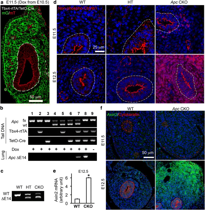
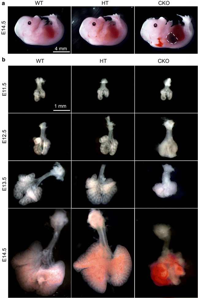
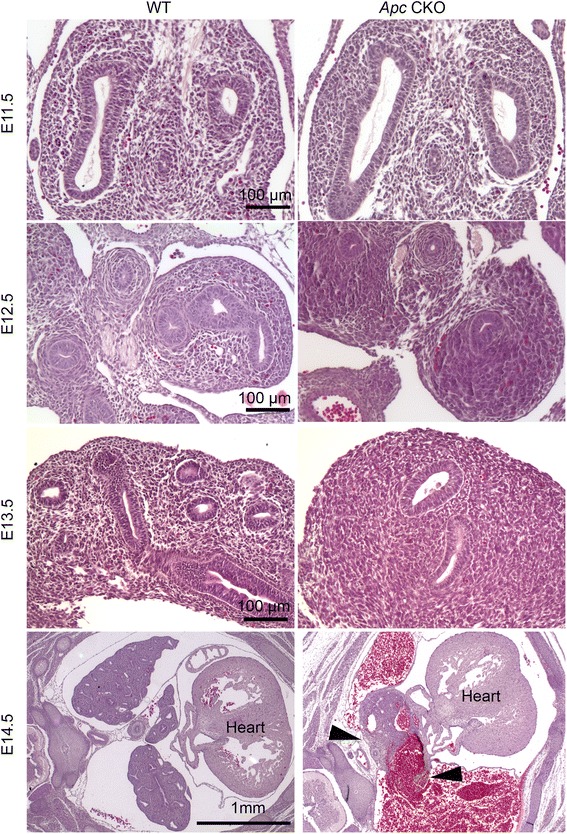
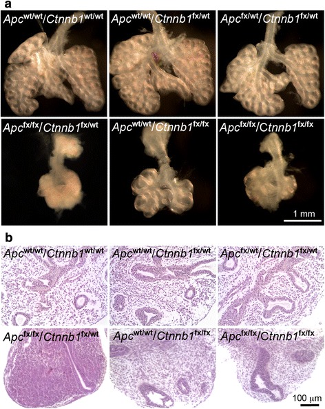
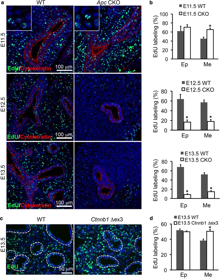
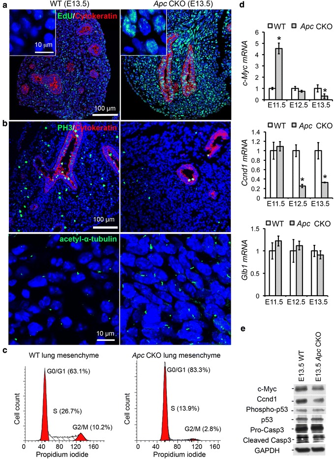
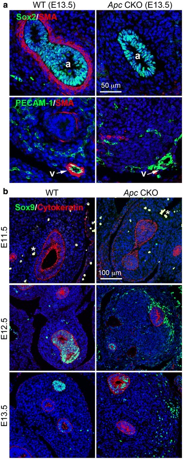
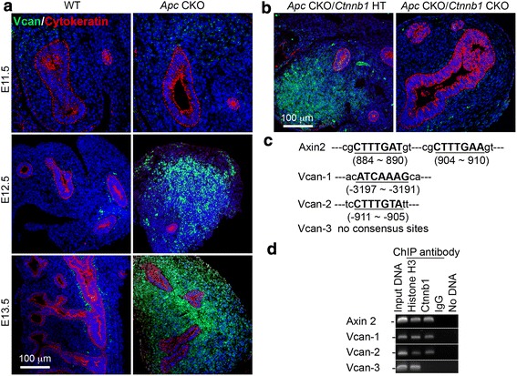
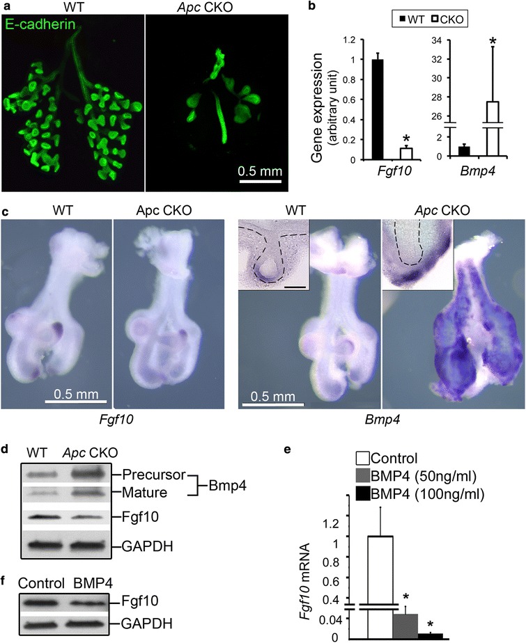
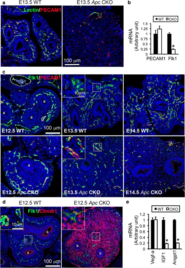
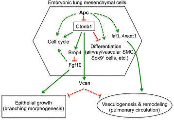
Results: We have specifically deleted the Apc gene in lung mesenchymal cells during early lung development in mice. Loss of Apc function resulted in immediate mesenchymal cell hyperproliferation through abnormal activation of Wnt/Ctnnb1, followed by a subsequent inhibition of cell proliferation due to cell cycle arrest at G0/G1, which was caused by a mechanism independent of Wnt/Ctnnb1. Meanwhile, abrogation of Apc also disrupted lung mesenchymal cell differentiation, including decreased airway and vascular smooth muscle cells, the presence of Sox9-positive mesenchymal cells in the peripheral lung, and excessive versican production. Moreover, lung epithelial branching morphogenesis was drastically inhibited due to disrupted Bmp4-Fgf10 morphogen production and regulation in surrounding lung mesenchyme. Lastly, lung mesenchyme-specific Apc conditional knockout also resulted in altered lung vasculogenesis and disrupted pulmonary vascular continuity through a paracrine mechanism, leading to massive pulmonary hemorrhage and lethality at mid-gestation when the pulmonary circulation should have started.

Conclusions: Our study suggests that Apc in lung mesenchyme plays central roles in coordinating the proper development of several quite different cellular compartments including lung epithelial branching and pulmonary vascular circulation during lung organogenesis.


Figures

Fig. 1
Fig. 1
Embryonic lung mesenchymal deletion of Apc induced ectopic activation of Wnt/Ctnnb1 signaling. a Induced Cre expression specifically in embryonic lung mesenchyme by a Tbx4 lung enhancer-driven Tet-On system was verified in an mT-mG reporter mouse lung (Tbx4-rtTA/TetO-Cre/mT-mG) one day after Dox administration (E10.5-E11.5). mG (green) was only expressed when upstream floxed-mT (red) was deleted by the induced Cre. b Lung specificity of Apc gene knockout at E11.5. Fetuses with different tail DNA genotypes were listed, and the heterozygous (lane 7) and homozygous (lane 8) deletions of floxed (fx)-exon 14 (ΔE14) were confirmed in lung tissue genomic DNAs. Leakage of Apc gene deletion in fetal lung was not detected in the absence of Dox induction (lane 9). c Truncation of Apc mRNA transcript in Apc conditional knockout lungs was verified by RT-PCR at E11.5. d Ectopic activation of Wnt/Ctnnb1 signaling in embryonic lung mesenchyme was detected by accumulated non-phospho (active) Ctnnb1 in both cytoplasm and nucleus of mesenchymal cells. Epithelial airway is highlighted with dashed line. e-f Increased Wnt canonical signaling activity in lung tissue was also verified by elevated Axin2 expression using real-time PCR (e; n = 5 in each group, *P < 0.05) and immunofluorescence staining (f)
Fig. 2
Fig. 2
Mesenchymal-specific deletion of Apc resulted in lung malformation. a Apc CKO induced from E10.5 led to thoracic hemorrhage indicated by dashed line at E14.5. b Gross view of isolated lungs showed that the Apc CKO caused mesenchymal hyperplasia at E12.5, growth arrest at E13.5, and massive hemorrhage at E14.5. No significant change was observed between WT and Apc heterozygous conditional knockout (HT) lungs
Fig. 3
Fig. 3
Dynamic changes of abnormal lung structure in Apc CKO lungs. There was no significant histological difference in E11.5 lungs between WT and Apc CKO mice. Aggregative growth of mesenchymal cells in Apc CKO lungs was apparent from E12.5. Lung tissue (arrowheads) was interspersed with massive areas of hemorrhage at E14.5
Fig. 4
Fig. 4
Phenotypic changes of E13.5 lungs with Apc and Ctnnb1 double conditional knockout in lung mesenchyme. Lung tissues with indicated Apc and Ctnnb1 genotypes were shown by gross view (a) and H&E-stained tissue sections (b). All fetal lungs shown here were positive for Tbx4-rtTA and TetO-Cre transgenes. Dox induction was started from E10.5
Fig. 5
Fig. 5
Dynamic changes in cell proliferation of Apc CKO lung. a-b Increased DNA synthesis, measured by EdU incorporation, in mesenchymal cells was found in lungs of Apc CKO mice at E11.5 (a-b), while decreased EdU labeling was observed in both epithelial and mesenchymal cells in Apc CKO lungs after E12.5. c-d Increased EdU incorporation persisted to E13.5 in lung mesenchyme when constitutively active Ctnnb1 (or Ctnnb1 Δex3) was expressed in mice with genotypes of Tbx4-Cre ERT/Ctnnb1 fx-ex3/+ plus tamoxifen induction at E10.5. a, c: Tissue immunofluorescence staining; b, d: quantitative analyses of the immunostained cells. Ep: epithelial cells, Me: mesenchymal cells. *P < 0.05, n = 4
Fig. 6
Fig. 6
Cell cycle arrest at G0/G1 phase in E13.5 Apc CKO lung. a Significant reduction of cell division in E13.5 Apc CKO lung, indicated by increased cells that retain EdU DNA labeling at E11.5. Increased nuclear size (insert) was apparent in Apc CKO lung. b Immunostaining for PH3 and acetyl-α-tubulin, markers for metaphase and G0/G1 phase, respectively. c FACS analysis of DNA contents for the cells isolated from E13.5 Apc CKO or WT control lungs. d Expression of key genes involved in regulation of cell cycle and senescence, detected by real-time PCR. *P < 0.05, n = 4. e Protein levels of the related genes were detected by western blot
Fig. 7
Fig. 7
Abrogation of Apc altered lung mesenchymal cell differentiation. a Deficient smooth muscle cell differentiation, detected by SMA immunostaining, was observed in both proximal airways (a) and vasculature (v) of E13.5 Apc CKO lung, which were marked by positively stained Sox2 and PECAM1, respectively. b Abnormal Sox9 expression was seen in the mesenchymal cells disseminated in peripheral lung of Apc CKO after E12.5. Lung epithelial cells were marked by cytokeratin staining. Cell nuclei were counterstained with DAPI (blue). *Hematopoietic cells with non-specific autofluorescence in E11.5 lungs
Fig. 8
Fig. 8
Hyperactivation of Wnt/Ctnnb1 pathway caused by Apc deletion in lung mesenchyme induced abnormal expression of Vcan. a Increased Vcan protein expression and distribution were detected in Apc CKO lung after E12.5, shown by immunofluorescence staining. Cell nuclei were counterstained with DAPI (blue). b Simultaneous deletion of Ctnnb1 in E13.5 Apc CKO lung mesenchyme blocked excessive Vcan expression. c Two sites containing Ctnnb1 binding DNA consensus sequences, which have been reported in Ctnnb1 target gene Axin2, were identified in Vcan promoter DNA. d Interaction between Ctnnb1 and Vcan promoter DNA was validated by ChIP using anti-active Ctnnb1 antibody. Anti-histone H3 and normal IgG were used as positive and negative controls, respectively
Fig. 9
Fig. 9
Lung epithelial branching morphogenesis was severely impaired in Apc CKO embryos. a Lung epithelial branches at E13.5 were visualized by whole mount E-cadherin immunofluorescence staining. b Altered expression of Fgf10 and Bmp4 in Apc CKO lung at E11.5 was detected by real-time PCR. *P < 0.05, n = 5. c Whole mount in situ hybridization showed that Fgf10 transcript was downregulated in the mesenchymal tips of Apc CKO lung at E11.5. In contrast, Bmp4 expression was dramatically increased in the sub-mesothelial mesenchyme of Apc CKO lung. Insert: distal tip in the vibratome section. Scale bar: 100 μm. d Alterations of Fgf10 and Bmp4 expression in E13.5 Apc CKO lung were verified at the protein level by western blot. e-f Treatment of cultured human fetal lung fibroblasts HLF1 with BMP4 (50–100 ng/ml) inhibited Fgf10 expression at the mRNA level (e, measured by real-time PCR) and the protein level (f, detected by western blot). *P < 0.05, n = 5
Fig. 10
Fig. 10
Apc knockout in lung mesenchyme disrupted pulmonary vascular network formation and pulmonary circulation continuity. a The endothelial cells of E13.5 pulmonary vasculature that was connected to the right heart were labeled with FITC-lectin after its intracardial injection, while all mature vascular endothelial cells were immunostained with anti-PECAM1. b Decreased expression of Flk1 at the mRNA level was readily detected in E12.5 Apc CKO lungs, *P < 0.05, n = 5. c Dynamic changes of angioblasts (Flk1+/PECAM1) and mature endothelial cells (Flk1+/PECAM1+) in Apc CKO lung. Reduced angioblasts were detected in Apc CKO lung at E13.5, while both angioblasts and mature endothelial cell numbers appeared significantly reduced in Apc CKO lung at E14.5. d Increased Ctnnb1 activation was not detected in Flk1+ cells in Apc CKO lung. e Reduced expression of Igf1 and Angpt1 at the mRNA level was detected in Apc CKO lungs at E12.5 (*P < 0.05, n = 5)
Fig. 11
Fig. 11
The postulated mechanisms of mesenchymal Apc as a central player in early lung development. Mesenchymal Apc not only regulates mesenchymal cell biology but also coordinates development of other cell compartments through both Wnt/Ctnnb1-dependent and Wnt/Ctnnb1-independent pathways

References

    1. Morrisey EE, Hogan BL. Preparing for the first breath: genetic and cellular mechanisms in lung development. Dev Cell. 2010;18:8–23. doi: 10.1016/j.devcel.2009.12.010. - DOI - PMC - PubMed
    1. de Mello DE, Sawyer D, Galvin N, Reid LM. Early fetal development of lung vasculature. Am J Respir Cell Mol Biol. 1997;16:568–81. doi: 10.1165/ajrcmb.16.5.9160839. - DOI - PubMed
    1. Groden J, Thliveris A, Samowitz W, Carlson M, Gelbert L, Albertsen H, et al. Identification and characterization of the familial adenomatous polyposis coli gene. Cell. 1991;66:589–600. doi: 10.1016/0092-8674(81)90021-0. - DOI - PubMed
    1. Aoki K, Taketo MM. Adenomatous polyposis coli (APC): a multi-functional tumor suppressor gene. J Cell Sci. 2007;120:3327–35. doi: 10.1242/jcs.03485. - DOI - PubMed
    1. Gurbuz AK, Giardiello FM, Petersen GM, Krush AJ, Offerhaus GJ, Booker SV, et al. Desmoid tumours in familial adenomatous polyposis. Gut. 1994;35:377–81. doi: 10.1136/gut.35.3.377. - DOI - PMC - PubMed

Publication types