. 2015 Aug;185(8):2232-45.
doi: 10.1016/j.ajpath.2015.04.016. Epub 2015 Jun 16.

The macrophage phagocytic receptor CD36 promotes fibrogenic pathways on removal of apoptotic cells during chronic kidney injury

Affiliations

The macrophage phagocytic receptor CD36 promotes fibrogenic pathways on removal of apoptotic cells during chronic kidney injury

Subramaniam Pennathur et al. Am J Pathol. 2015 Aug.

Abstract

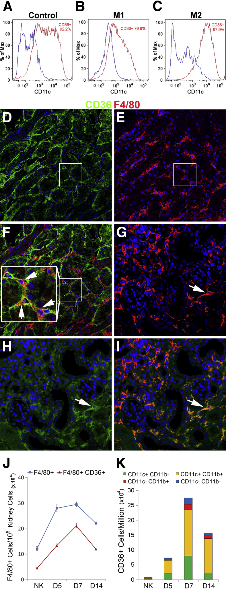
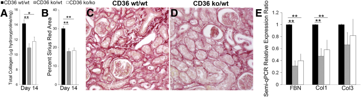
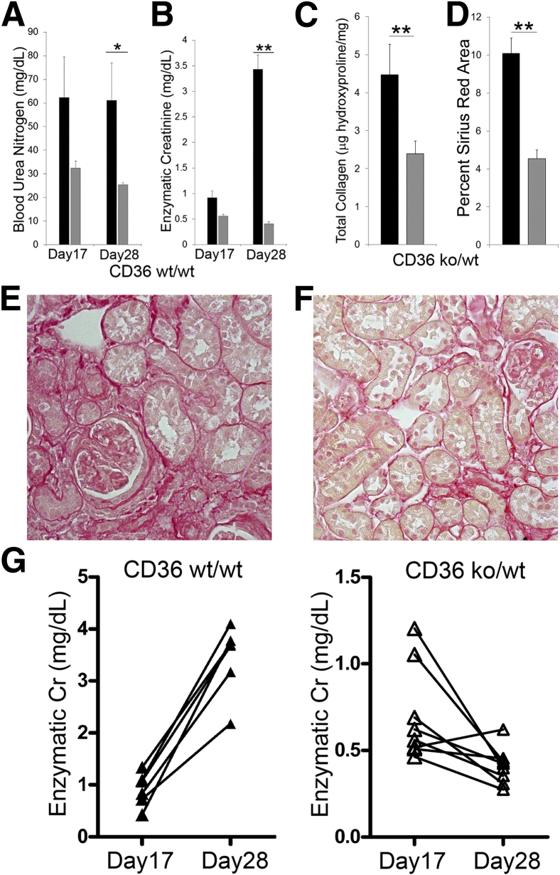
The removal of apoptotic cells is an innate function of tissue macrophages; however, its role in disease progression is unclear. The present study was designed to investigate the role of macrophage CD36, a recognized receptor of apoptotic cells and oxidized lipids, in two models of kidney injury: unilateral ureteral obstruction (UUO) and ischemia reperfusion. To differentiate the macrophage CD36-specific effects in vivo, we generated CD36 chimeric mice by bone marrow transplantation and evaluated the two models. Fibrosis severity was substantially decreased after UUO with a corresponding decrease in matrix synthesis in macrophage CD36-deficient mice. Despite a reduction in fibrosis severity, a 56% increase in apoptotic cells was found without an increase in apoptotic effectors. In addition, a substantial reduction was observed in tumor necrosis factor-α and transforming growth factor-β1 mRNA levels and intracellular bioactive oxidized lipid levels in CD36-deficient macrophages. To validate the functional role of macrophage CD36, we performed unilateral ischemia reperfusion, followed by contralateral nephrectomy. Similarly, we found that the severity of fibrosis was reduced by 55% with a corresponding improvement in kidney function by 88% in macrophage CD36-deficient mice. Taken together, these data suggest that macrophage CD36 is a critical regulator of oxidative fibrogenic signaling and that CD36-mediated phagocytosis of apoptotic cells may serve as an important pathway in the progression of fibrosis.


Figures

Figure 1
Figure 1
Scavenger receptor CD36 identifies a phagocytic M2 macrophage. Wild-type BMDMs were activated with M1 (LPS/IFNγ) or M2 (IL-4) stimuli. BMDMs (CD45+ CD11c+) were analyzed for CD36 expression by FACS (blue CD36, red CD36+) at baseline (A) and at M1 (B) and M2 (C) activation. Representative confocal images in normal kidneys: CD36 (green; D, boxed areas), F4/80 (red; E, boxed areas), and merged (F, inset); arrows in inset indicate CD36+ resident macrophages surrounding renal tubules. After UUO, a significant portion of the F4/80+ population (G) are CD36+ (H) and merged (I). Arrows indicate a subpopulation of CD36+ phagocytic macrophages (GI). J: Graph summarizes FACS analysis of CD45+F4/80+ subpopulations by CD36 expression. K: Graph summarizes FACS analysis of CD45+CD36+ subpopulations from UUO kidneys at day 5, 7, and 14 by the markers CD11b and CD11c (Supplemental Figure S2). Data are expressed as means ± SEM. n = 4 per group. Original magnification: ×400 (DF). BMDM, bone marrow-derived macrophage; Cy5.5, cyanine 5.5; FACS, fluorescent-activated cell sorting; IFN, interferon; LPS, lipopolysaccharide; M1, classically activated; M2, alternatively activated; NK, contralateral kidney; PerCP, peridinin chlorophyll; UUO, unilateral ureteral obstruction.
Figure 2
Figure 2
CD36-dependent apoptotic cell clearance activates fibrogenic pathways. Apoptotic tubular cells were generated by irradiating MCT cells. A and B: FACS analysis with Annexin V and propidium iodide confirm late-stage apoptosis in MCT cells. Mouse peritoneal TEMs were co-cultured with apoptotic MCT cells labeled with CellTracker (red). C: Arrow indicates CD36+/+ TEM engulfing Annexin V+ MCT cells. D: Apoptotic cells are cleared at a reduced rate in CD36−/− TEMs. Arrow indicates apoptotic cell bound to CD36−/− TEMs but not engulfed. Semiquantitative real-time PCR was performed 24 hours after co-culture of TEMs and apoptotic MCT cells. Graphs summarize expression levels normalized to 18S and GAPDH: TNFα (E), TNFα receptor 1 (F), TGF-β1 (G), and TGF-β1 receptor (H). Comparison groups are CD36+/+ mphi only (ac−) versus CD36+/+ mphi with ac; and CD36+/+ mphi with ac versus CD36−/− mphi with ac. Data are expressed as means ± SEM. n = 4 per group; P < 0.05, ∗∗P < 0.01. Original magnification: ×800 (C and D). ac, apoptotic cell; FACS, fluorescent-activated cell sorting; FITC, fluorescein isothiocyanate; MCT, mouse cortical tubular; mphi, TEM; TEM, thioglycollate-elicited macrophage; TGF, transforming growth factor; TNF, tumor necrosis factor.
Figure 3
Figure 3
Macrophage CD36 is a profibrotic phenotype. A: Total kidney collagen content, measured with the hydroxyproline assay. B: The graph summarizes the results of picrosirius red+ interstitial collagen quantification, analyzed by nested analysis of variance, with representative images (C and D). E: The graph shows the results of analysis of kidney extracellular matrix mRNA levels, measured by semi-qPCR and normalized to two housekeeping genes, 18S and GAPDH: FBN, Col1, and Col3. Data are expressed as means ± SEM. n = 5 to 6 per group. P < 0.05, ∗∗P < 0.01. Original magnification: ×400 (C and D). Col1, procollagen I; Col3, procollagen III; FBN, fibronectin; KO, knockout; semi-qPCR, semiquantitative real-time PCR; wt, wild-type.
Figure 4
Figure 4
Macrophage CD36 clears apoptotic cells during chronic kidney injury. Apoptotic cells were identified by TUNEL staining in day 14 UUO kidneys. A and B: Representative images with arrows that denote TUNEL+ apoptotic cells. C: Graph summarizes analysis by U test. Data are expressed as means ± SEM. n = 5 to 6 per group. P < 0.05, ∗∗P < 0.01. Original magnification, ×400. ko, knockout; TUNEL, terminal deoxynucleotidyl transferase-mediated dUTP nick-end labelling; UUO, unilateral ureteral obstruction; wt, wild-type.
Figure 5
Figure 5
Macrophage CD36 promotes tubular injury. PAS-stained sections were analyzed for tubular injury. AC: Representative images from day 14 UUO kidneys of CD36 chimeric mice. D: Graph summarizes the results of PAS tubular injury scores analyzed by U test. Lcn2 is a marker of renal tubular injury. Representative images of Lcn2 from low power (E–G) and high power (HJ) in chimeric mice. Boxed areas indicate area of high power image. K: Graph summarizes semiquantitative image analysis of Lcn2 staining by nested analysis of variance. Solid bar representsCD36wt/wt; gray bar, CD36ko/wt; open bar, CD36ko/ko. Data are expressed as means ± SEM. n = 5 to 6 per group (D); n = 6 to 7 per group (K). ∗∗P < 0.01. Original magnification: ×400 (AC); ×100 (E–G). ko, knockout; Lcn2, Lipocalin 2; PAS, periodic acid-Schiff; UUO, unilateral ureteral obstruction; wt, wild-type.
Figure 6
Figure 6
Macrophage CD36 activates fibrogenic signaling pathways. A: Representative Western blot analysis of phosphorylated IκB-α and total IκB-α from total kidney homogenate of day 14 UUO kidneys. B: Graph summarizes results of quantification of band densities of phosphorylated IκB-α and total IκB-α ratios. Proinflammatory and profibrotic mRNA levels in total RNA from whole kidney were measured by semi-qPCR and were normalized to two housekeeping genes, 18S and GAPDH. C: The graph summarizes the results on day 14 UUO kidneys from CD36 chimeric mice. CD11b+ cells were isolated from day14 UUO kidneys of CD36+/+ and CD36−/− mice, and total RNA was analyzed by semi-qPCR normalized to 18S and GAPDH. D: Graph summarizes the mRNA expression levels of proinflammatory and profibrotic cytokines. CD36 co-immunoprecipitation was performed on a pooled sample of DSP-treated, CD11b+ isolated cells from day 7 UUO kidneys to determine intracellular signaling binding partners. Co-immunoprecipitation proteins were eluted from CD36 antibody beads, and Western blot analysis was performed. E: Representative Western blot analysis probed for JNK2 and Lyn kinase. Data are expressed as means ± SEM. n = 5 to 6 per group. P < 0.05, ∗∗P < 0.01. DSP, dithiobis(succinimidyl propionate); IκB-α, nuclear factor of κ light polypeptide gene enhancer in B-cells inhibitor α; JNK2, c-Jun N-terminal kinase-2; ko, knockout; semi-qPCR, semiquantitative real-time PCR; TGF, transforming growth factor; TNF, tumor necrosis factor; UUO, unilateral ureteral obstruction; wt, wild-type.
Figure 7
Figure 7
Macrophage bioactive lipids are reduced in the absence of CD36. Interstitial macrophages (CD11b+) were isolated from contralateral (NK) and UUO day 14 kidneys from CD36+/+ and CD36−/− mice. Samples were analyzed by reverse-phase HPLC and mass spectrometry for 5-, 12-, 15-, 20-HETE (AD) and 9S-, 13S-HODE (E and F). Concentrations were normalized to their precursor: AA for HETEs and LA for HODEs. Black bar represents CD36+/+; white bar, CD36−/−. Data are expressed as means ± SEM. n = 4 to 5 per group. P < 0.05, ∗∗P < 0.01. AA, arachidonic acid; HETE, hydroxyeicosatetraenoic acid; HODE, hydroxyoctadecadienoic acid; HPLC, high-performance liquid chromatography; LA, linoleic acid; NK, normal kidney; UUO, unilateral ureteral obstruction.
Figure 8
Figure 8
Absence of CD36 macrophages improves kidney function after ischemic injury. Unilateral IR was performed, and the contralateral kidney was surgically removed at day 14 after IR. Phlebotomy was performed at day 17 and 28 (sacrifice). Graphs summarize the results of BUN (A) and enzymatic creatinine (B) concentrations from CD36 chimeric mice. Total kidney collagen content was measured by hydroxyproline assay and summarized in graph C. D: Graph summarizes image analysis of picrosirius red+ interstitial collagen quantification by nested analysis of variance, with representative images (E and F). The fold change (δ) in enzymatic creatinine from day 17 to day 28 was calculated and analyzed by Student's t-test. G: Graphs demonstrate the change in Cr in CD36 chimeric mice (mean fold change: CD36wt/wt = δ3.3 ± 0.9 mg/dL versus CD36ko/wt = δ−0.4 ± 0.1 mg/dL). Black bar representsCD36wt/wt; gray bar, CD36ko/wt. Data are expressed as means ± SEM. n = 6 to 8 per group. P < 0.05, ∗∗P < 0.01. Original magnification, ×400. BUN, blood urea nitrogen; Cr, creatinine; IR, ischemia reperfusion; KO, knockout; wt, wild-type.
Figure 9
Figure 9
Absence of CD36 macrophages reduces tubular injury. PAS-stained sections were analyzed for tubular injury at day 28 after IR. A and B: Representative images of IR kidneys of CD36 chimeric mice. C: Graph summarizes the results of PAS tubular injury scores analyzed by U test. D and E: Representative images of Lcn2 from low power. F: Graph summarizes semiquantitative image analysis of Lcn2 staining by nested analysis of variance and high power (G and H) in chimeric mice. Boxed areas in D and E indicate area of high-power image. Proinflammatory and profibrotic mRNA levels in total RNA from whole kidney were measured by semiquantitative real-time PCR and normalized to two housekeeping genes, 18S and GAPDH. I: The graph summarizes the results on day 28 IR kidneys from CD36 chimeric mice. Black bar representsCD36wt/wt; gray bar, CD36ko/wt; white bar, sham; Data are expressed as means ± SEM. n = 5 to 6 per group (C); n = 7 to 8 per group (F); n = 6 per group (I). ∗∗P < 0.01. Original magnification: ×400 (A and B). IR, ischemia reperfusion; ko, knockout; Lcn2, Lipocalin 2; PAS, periodic acid-Schiff; semi-qPCR, semiquantitative real-time PCR; wt, wild-type.

References

    1. Wahl S.M., McCartney-Francis N., Allen J.B., Dougherty E.B., Dougherty S.F. Macrophage production of TGF-beta and regulation by TGF-beta. Ann N Y Acad Sci. 1990;593:188–196. - PubMed
    1. Bonner J.C., Osornio-Vargas A.R., Badgett A., Brody A.R. Differential proliferation of rat lung fibroblasts induced by the platelet-derived growth factor-AA, -AB, and -BB isoforms secreted by rat alveolar macrophages. Am J Respir Cell Mol Biol. 1991;5:539–547. - PubMed
    1. Duffield J.S., Forbes S.J., Constandinou C.M., Clay S., Partolina M., Vuthoori S., Wu S., Lang R., Iredale J.P. Selective depletion of macrophages reveals distinct, opposing roles during liver injury and repair. J Clin Invest. 2005;115:56–65. - PMC - PubMed
    1. Duffield J.S., Tipping P.G., Kipari T., Cailhier J.F., Clay S., Lang R., Bonventre J.V., Hughes J. Conditional ablation of macrophages halts progression of crescentic glomerulonephritis. Am J Pathol. 2005;167:1207–1219. - PMC - PubMed
    1. Martinez F.O., Gordon S., Locati M., Mantovani A. Transcriptional profiling of the human monocyte-to-macrophage differentiation and polarization: new molecules and patterns of gene expression. J Immunol. 2006;177:7303–7311. - PubMed

Publication types

LinkOut - more resources