

Using Ambystoma mexicanum (Mexican axolotl) embryos, chemical genetics, and microarray analysis to identify signaling pathways associated with tissue regeneration
- PMID: 26092703
- PMCID: PMC4662883
- DOI: 10.1016/j.cbpc.2015.06.004
Using Ambystoma mexicanum (Mexican axolotl) embryos, chemical genetics, and microarray analysis to identify signaling pathways associated with tissue regeneration
Abstract
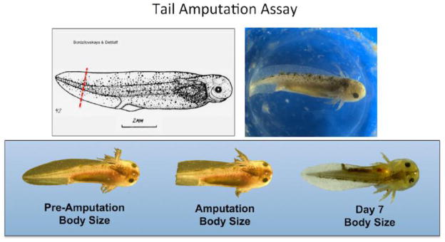
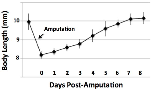
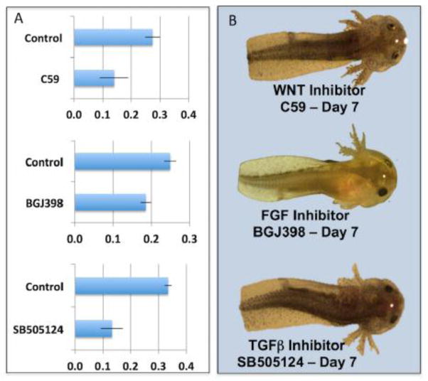
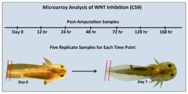
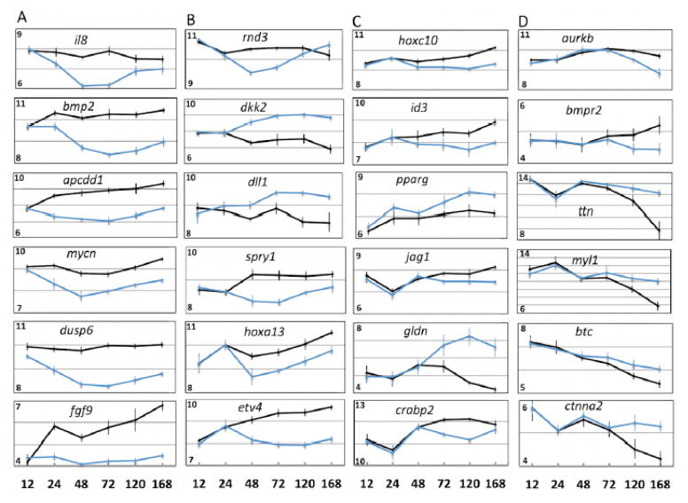
Amphibian vertebrates are important models in regenerative biology because they present exceptional regenerative capabilities throughout life. However, it takes considerable effort to rear amphibians to juvenile and adult stages for regeneration studies, and the relatively large sizes that frogs and salamanders achieve during development make them difficult to use in chemical screens. Here, we introduce a new tail regeneration model using late stage Mexican axolotl embryos. We show that axolotl embryos completely regenerate amputated tails in 7days before they exhaust their yolk supply and begin to feed. Further, we show that axolotl embryos can be efficiently reared in microtiter plates to achieve moderate throughput screening of soluble chemicals to investigate toxicity and identify molecules that alter regenerative outcome. As proof of principle, we identified integration 1 / wingless (Wnt), transforming growth factor beta (Tgf-β), and fibroblast growth factor (Fgf) pathway antagonists that completely block tail regeneration and additional chemicals that significantly affected tail outgrowth. Furthermore, we used microarray analysis to show that inhibition of Wnt signaling broadly affects transcription of genes associated with Wnt, Fgf, Tgf-β, epidermal growth factor (Egf), Notch, nerve growth factor (Ngf), homeotic gene (Hox), rat sarcoma/mitogen-activated protein kinase (Ras/Mapk), myelocytomatosis viral oncogene (Myc), tumor protein 53 (p53), and retinoic acid (RA) pathways. Punctuated changes in the expression of genes known to regulate vertebrate development were observed; this suggests the tail regeneration transcriptional program is hierarchically structured and temporally ordered. Our study establishes the axolotl as a chemical screening model to investigate signaling pathways associated with tissue regeneration.
Keywords: Axolotl; C59; Chemical screening; Regeneration; Wnt.
Copyright © 2015 Elsevier Inc. All rights reserved.
Figures
References
-
- Bordzilovskaya NP, Dettlaff TA, Duhon ST, Malacinski GM. Developmental-stage series of axolotl embryos. In: Armstrong JB, Malacinski GM, editors. Developmental Biology of the Axolotl. Oxford University Press; New York: 1989. pp. 201–219.
-
- Charlemagne J. Morphological studies of blood cell differentiation in the axolotl, Ambystoma mexicanum Shaw. Z Zellforsch Mikrosk Anat. 1972;123:224–239. - PubMed
-
- Chen Y, Love NR, Amaya E. Tadpole tail regeneration in Xenopus. Biochem Soc Trans. 2014;42:617–623. - PubMed
-
- Fellah JS, Vaulot D, Tournefier A, Charlemagne J. Ontogeny of immunoglobulin expression in the Mexican axolotl. Development. 1989;107:253–263. - PubMed
Publication types
MeSH terms
Grants and funding
LinkOut - more resources
Full Text Sources
Other Literature Sources
Research Materials
Miscellaneous
