Practice Guideline
. 2015 Jul 14;85(2):177-89.
doi: 10.1212/WNL.0000000000001729. Epub 2015 Jun 19.

International consensus diagnostic criteria for neuromyelitis optica spectrum disorders

Affiliations
Practice Guideline

International consensus diagnostic criteria for neuromyelitis optica spectrum disorders

Dean M Wingerchuk et al. Neurology. .

Abstract

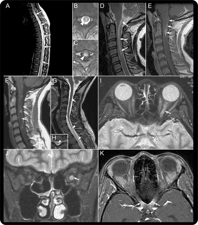
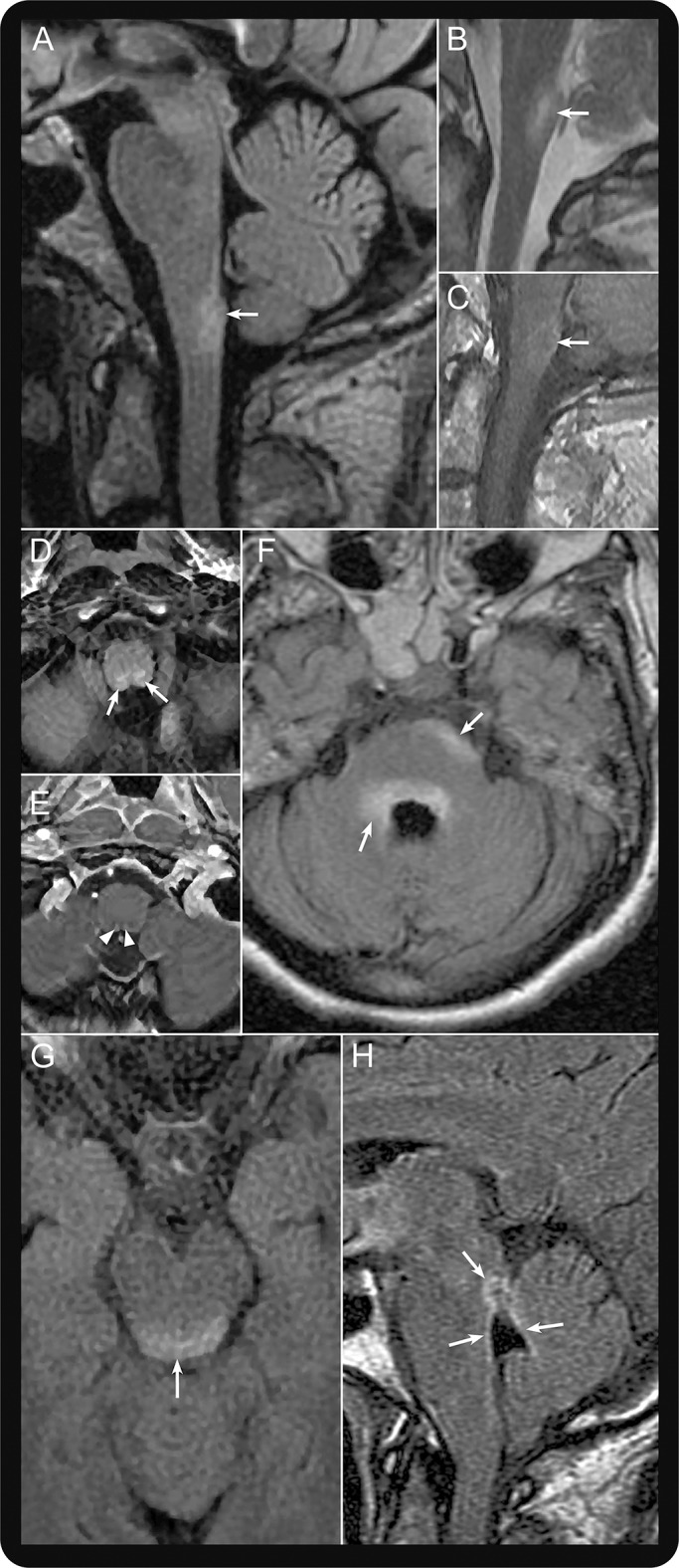
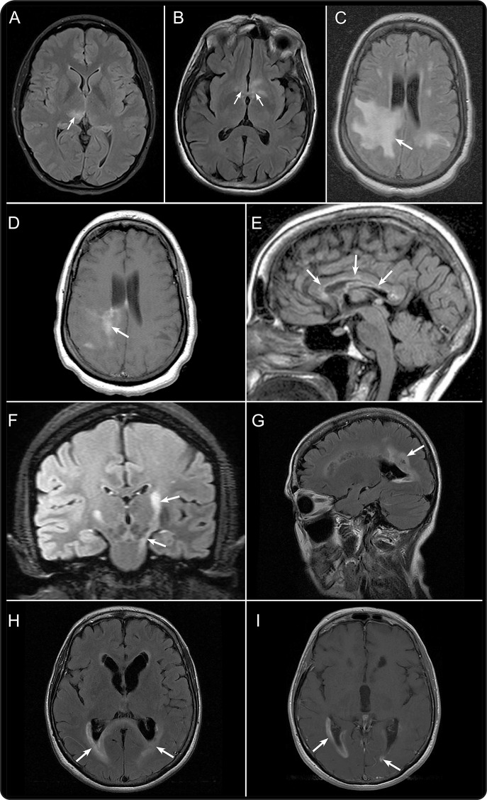
Neuromyelitis optica (NMO) is an inflammatory CNS syndrome distinct from multiple sclerosis (MS) that is associated with serum aquaporin-4 immunoglobulin G antibodies (AQP4-IgG). Prior NMO diagnostic criteria required optic nerve and spinal cord involvement but more restricted or more extensive CNS involvement may occur. The International Panel for NMO Diagnosis (IPND) was convened to develop revised diagnostic criteria using systematic literature reviews and electronic surveys to facilitate consensus. The new nomenclature defines the unifying term NMO spectrum disorders (NMOSD), which is stratified further by serologic testing (NMOSD with or without AQP4-IgG). The core clinical characteristics required for patients with NMOSD with AQP4-IgG include clinical syndromes or MRI findings related to optic nerve, spinal cord, area postrema, other brainstem, diencephalic, or cerebral presentations. More stringent clinical criteria, with additional neuroimaging findings, are required for diagnosis of NMOSD without AQP4-IgG or when serologic testing is unavailable. The IPND also proposed validation strategies and achieved consensus on pediatric NMOSD diagnosis and the concepts of monophasic NMOSD and opticospinal MS.


Figures

Figure 1
Figure 1. Spinal cord and optic nerve MRI patterns in neuromyelitis optica spectrum disorder
Spinal cord imaging in the context of acute myelitis in neuromyelitis optica spectrum disorders (NMOSD) usually reveals a longitudinally extensive transverse myelitis (LETM) lesion extending over 3 or more vertebral segments. Sagittal T2-weighted MRI of the thoracic spinal cord (A) demonstrates a typical LETM lesion involving most of the thoracic spinal cord (arrows). LETM lesions have a predilection for the central cord, as shown by axial T2-weighted (B; arrowhead) and T1-weighted MRI with gadolinium (C; arrowhead). Cervical LETM may extend into the medulla, a characteristic NMOSD pattern demonstrated in D (arrows; sagittal T2-weighted MRI) and E (arrows; sagittal T1-weighted MRI with gadolinium). Acute LETM lesions can be associated with intralesional hypointensity as shown by sagittal T1-weighted MRI (F; arrow); in this example, a rim of gadolinium enhancement surrounds the hypointense region. Chronic sequelae of LETM may include longitudinally extensive segments of spinal cord atrophy as shown by T2-weighted MRI using sagittal (G; the 2 arrowheads indicate the atrophic segment and the top arrow indicates the normal diameter of unaffected cervical spinal cord) and axial planes (H; arrowhead shows an atrophic spinal cord). Fast spin echo fat-suppressed T2-weighted MRI in the axial (I) and coronal (J) planes shows increased signal throughout most the length of the left optic nerve, especially its posterior portion (arrows). Axial T1-weighted MRI with gadolinium shows enhancement of the optic chiasm (K; arrows). These images are from 2 different patients experiencing acute optic neuritis in the setting of NMOSD.
Figure 2
Figure 2. Dorsal medulla, area postrema, and other brainstem lesions in neuromyelitis optica spectrum disorder
Sagittal T2-weighted fluid-attenuated inversion recovery (FLAIR) MRI shows a lesion in the dorsal medulla (A; arrow). Sagittal T2-weighted (B) and T1-weighted MRI with gadolinium (C) each demonstrate an acute lesion (arrows) associated with area postrema clinical syndrome. Axial T2-weighted FLAIR (D; arrows) and T1-weighted MRI with gadolinium (E; arrowheads) show dorsal medulla involvement in a patient with acute area postrema clinical syndrome. Axial T2-weighted FLAIR MRI shows periependymal lesions involving the pons (F; arrows) and dorsal midbrain (G; arrow). Sagittal T2-weighted FLAIR MRI shows increased signal surrounding the fourth ventricle (H; arrows).
Figure 3
Figure 3. Diencephalic and cerebral lesions in neuromyelitis optica spectrum disorder
A variety of brain lesion patterns are associated with neuromyelitis optica spectrum disorder. Axial T2-weighted fluid-attenuated inversion recovery (FLAIR) MRI from 2 patients demonstrates lesions involving the right thalamus (A; arrow) and the hypothalamus (B; arrows). Axial T2-weighted FLAIR MRI shows an extensive subcortical white matter lesion (C; arrow) that enhances after gadolinium administration on T1-weighted sequences (D; arrow). Chronic longitudinally extensive and linear corpus callosum lesions are depicted on sagittal T2-weighted FLAIR MRI (E; arrows). Coronal T2-weighted FLAIR MRI shows longitudinal involvement of the corticospinal tract extending to the cerebral peduncle and pons (F; arrows). Acute periependymal cerebral lesions from one patient are depicted using sagittal (G; arrow) and axial (H; arrows) T2-weighted FLAIR MRI and axial T1-weighted MRI with gadolinium (I; arrows).

Comment in

References

    1. Wingerchuk DM, Lennon VA, Lucchinetti CF, Pittock SJ, Weinshenker BG. The spectrum of neuromyelitis optica. Lancet Neurol 2007;6:805–815. - PubMed
    1. Wingerchuk DM, Lennon VA, Pittock SJ, Lucchinetti CF, Weinshenker BG. Revised diagnostic criteria for neuromyelitis optica. Neurology 2006;66:1485–1489. - PubMed
    1. Jarius S, Wildemann B. The history of neuromyelitis optica. J Neuroinflammation 2013;10:8. - PMC - PubMed
    1. Wingerchuk DM, Hogancamp WF, O'Brien PC, Weinshenker BG. The clinical course of neuromyelitis optica (Devic's syndrome). Neurology 1999;53:1107–1114. - PubMed
    1. Lennon VA, Wingerchuk DM, Kryzer TJ, et al. A serum autoantibody marker of neuromyelitis optica: distinction from multiple sclerosis. Lancet 2004;364:2106–2112. - PubMed

Publication types