. 2015 Jun 21:16:154.
doi: 10.1186/s12891-015-0615-1.

α-Melanocyte-stimulating-hormone (α-MSH) modulates human chondrocyte activation induced by proinflammatory cytokines

Affiliations

α-Melanocyte-stimulating-hormone (α-MSH) modulates human chondrocyte activation induced by proinflammatory cytokines

Franco Capsoni et al. BMC Musculoskelet Disord. .

Abstract

Background: Alpha-melanocyte-stimulating-hormone (α-MSH) has marked anti-inflammatory potential. Proinflammatory cytokines are critical mediators of the disturbed cartilage homeostasis in osteoarthritis, inhibiting anabolic activities and increasing catabolic activities in chondrocytes. Since human chondrocytes express α-MSH receptors, we evaluated the role of the peptide in modulating chondrocyte production of pro-inflammatory cytokines, matrix metalloproteinases (MMPs), tissue inhibitors of MMPs (TIMPs), inducible nitric oxide synthase (iNOS) and nitric oxide (NO) in response to interleukin-1β (IL-1β) and tumor necrosis factor-α (TNF-α).

Methods: Human articular chondrocytes were obtained from osteoarthritic joint cartilage from subjects undergoing hip routine arthroplasty procedures. The cells were cultured with or without α-MSH in the presence of IL-1β or TNF-α. Cell-free supernatants were collected and cells immediately lysed for RNA purification. Expression of cytokines, MMPs, TIMPs, iNOS was determined by Reverse Transcription Real-time Polymerase Chain Reaction and enzyme-linked immunosorbent assay. Griess reaction was used for NO quantification.

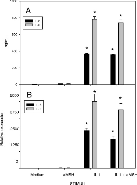
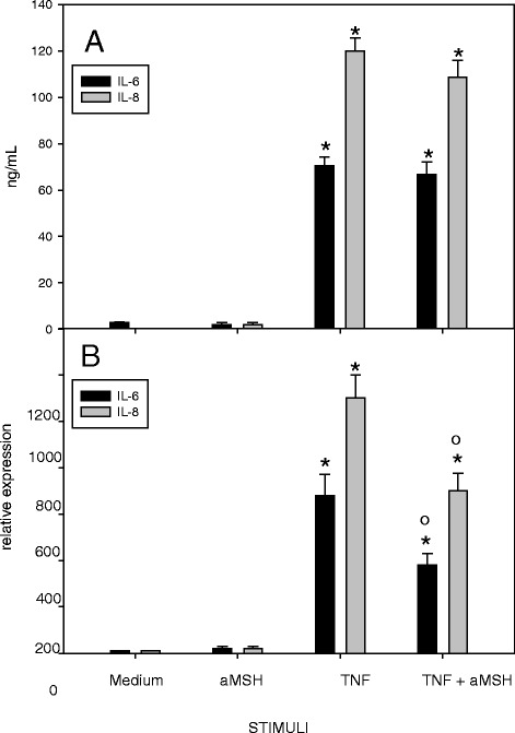
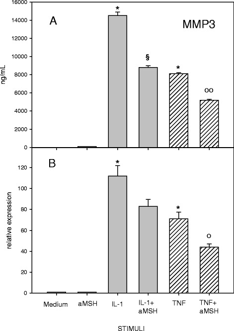
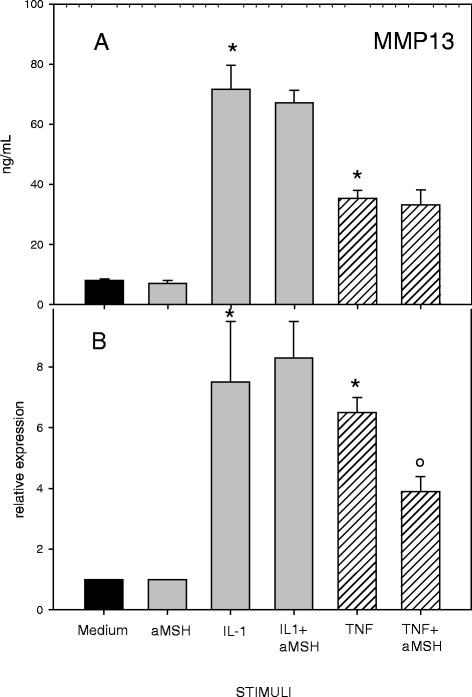
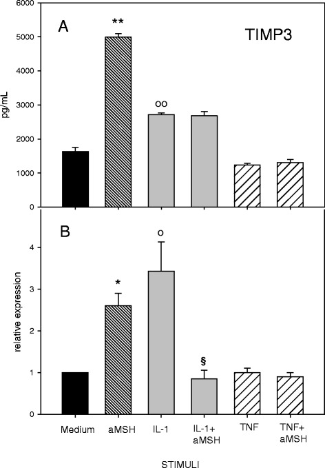
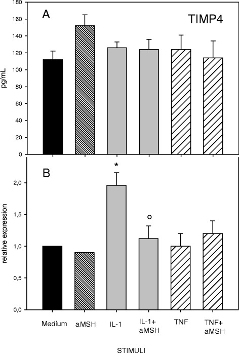
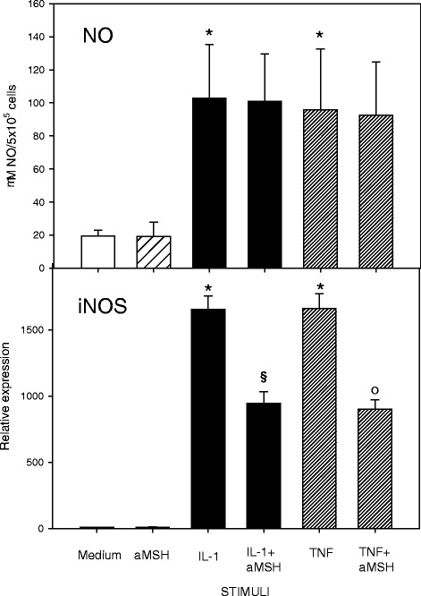
Results: Gene expression and secretion of IL-6, IL-8, MMP-3, MMP-13 were significantly increased in IL-1β or TNF-α-stimulated chondrocytes; α-MSH did not modify the release of IL-6 or IL-8 while the peptide significantly reduced their gene expression on TNF-α-stimulated cells. A significant inhibition of MMP3 gene expression and secretion from IL-1β or TNFα-stimulated chondrocytes was induced by α-MSH. On the other hand, α-MSH did not modify the release of MMP-13 by cytokine-stimulated chondrocyte but significantly decreased gene expression of the molecule on TNF-α-stimulated cells. Detectable amount of TIMP-3 and TIMP-4 were present in the supernatants of resting chondrocytes and a significant increase of TIMP-3 gene expression and release was induced by α-MSH on unstimulated cells. TIMP-3 secretion and gene expression were significantly increased in IL-1β-stimulated chondrocytes and α-MSH down-regulated gene expression but not secretion of the molecule. TIMP-4 gene expression (but not secretion) was moderately induced in IL-1β-stimulated chondrocytes with a down-regulation exerted by α-MSH. IL-1β and TNF-α were potent stimuli for NO production and iNOS gene expression by chondrocytes; no inhibition was induced by α-MSH on cytokine-stimulated NO production, while the peptide significantly reduced gene expression of iNOS.

Conclusions: Our results underscore a potential anti-inflammatory and chondroprotective activity exerted by α-MSH, increasing TIMP-3 gene expression and release on resting cells and down- modulating TNF-α-induced activation of human chondrocytes. However, the discrepancy between the influences exerted by α-MSH on gene expression and protein release as well as the difference in the inhibitory pattern exerted by α-MSH in TNF-α- or IL-1β-stimulated cells leave some uncertainty on the role of the peptide on chondrocyte modulation.


Figures

Fig. 1
Fig. 1
Effect of α-MSH on IL-6 and IL-8 secretion (a) and gene expression (b) in IL-1β-stimulated chondrocytes. Human primary chondrocytes were incubated for 4 h at 37 °C with 10−6M α-MSH and further cultured for 40 h in the presence or absence of 10 ng/mL IL-1β. IL-6 and IL-8 concentration in the supernatans (a) and gene expression (b) were then measured. Data from 4 separate experiments (± S.E.M.) are shown. *p < 0.0001 vs Medium
Fig. 2
Fig. 2
Effect of α-MSH on IL-6 and IL-8 secretion (a) and gene expression (b) in TNF-α-stimulated chondrocytes. Human primary chondrocytes were incubated for 4 h at 37 °C with 10−6M α-MSH and further cultured for 40 h in the presence or absence of 10 ng/mL TNF-α. IL-6 and IL-8 concentration in the supernatans (a) and gene expression (b) were then measured. Data from 4 separate experiments (± S.E.M.) are shown. *p < 0.0001 vs Medium; ° p < 0.05 vs TNF-α
Fig. 3
Fig. 3
Effect of α-MSH on MMP-3 secretion (a) and gene expression (b) in IL-1β or TNF-α-stimulated chondrocytes. Human primary chondrocytes were incubated for 4 h at 37 °C with 10−6M α-MSH and further cultured for 40 h in the presence or absence of 10 ng/mL IL-1β or TNF-α. MMP-3 concentration in the supernatans (a) and gene expression (b) were then measured. Data from 4 separate experiments (± S.E.M.) are shown. * p < 0.0001 vs Medium; § p < 0.001 vs IL-1β; ° p < 0.01 vs TNF-α; °° p < 0.001 vs TNF-α
Fig. 4
Fig. 4
Effect of α-MSH on MMP-13 secretion (a) and gene expression (b) in IL-1β or TNF-α-stimulated chondrocytes. Human primary chondrocytes were incubated for 4 h at 37 °C with 10−6M α-MSH and further cultured for 40 h in the presence or absence of 10 ng/mL IL-1β or TNF-α. MMP-13 concentration in the supernatans (a) and gene expression (b) were then measured. Data from 4 separate experiments (± S.E.M.) are shown. * p < 0.0001 vs Medium; °p < 0.05 vs TNF-α
Fig. 5
Fig. 5
Effect of α-MSH on TIMP-3 secretion (a) and gene expression (b) in IL-1β or TNF-α-stimulated chondrocytes. Human primary chondrocytes were incubated for 4 h at 37 °C with 10−6M α-MSH and further cultured for 40 h in the presence or absence of 10 ng/mL IL-1β or TNF-α. TIMP-3 concentration in the supernatans (a) and gene expression (b) were then measured. Data from 4 separate experiments (± S.E.M.) are shown. *P < 0.002 vs Medium; **p < 0.001 vs Medium; ° p < 0.02 vs Medium; °° p < 0.002 vs Medium; § p < 0.05 vs IL-1β
Fig. 6
Fig. 6
Effect of α-MSH on TIMP-4 secretion (a) and gene expression (b) in IL-1β or TNF-α-stimulated chondrocytes. Human primary chondrocytes were incubated for 4 h at 37 °C with 10−6M α-MSH and further cultured for 40 h in the presence or absence of 10 ng/mL IL-1β or TNF-α. TIMP-4 concentration in the supernatans (a) and gene expression (b) were then measured. Data from 4 separate experiments (± S.E.M.) are shown. *p < 0.02 vs Medium; ° P < 0.05 vs IL-1β
Fig. 7
Fig. 7
Effect of α-MSH on NO secretion and iNOS gene expression in IL-1β or TNF-α-stimulated chondrocytes. Human primary chondrocytes were incubated for 4 h at 37 °C with 10−6M α-MSH and further cultured for 40 h in the presence or absence of 10 ng/mL IL-1β or TNF-α. NO concentration in the supernatans (a) and iNOS gene expression (b) were then measured. Data from 4 separate experiments (± S.E.M.) are shown. *p < 0.0001 vs Medium; § p < 0.005 vs IL-1β; °p < 0.005 vs TNF-α

References

    1. Goldring MB. Update on the biology of the chondrocyte and new approaches to treating cartilage diseases. Best Pract Res Clin Rheumatol. 2006;20:1003–25. doi: 10.1016/j.berh.2006.06.003. - DOI - PubMed
    1. Aigner T, Sachse A, Gebhard PM, Roach HI. Osteoarthritis: Pathobiology-targets and ways for therapeutic intervention. Adv Drug Deliv Rev. 2006;58:128–49. doi: 10.1016/j.addr.2006.01.020. - DOI - PubMed
    1. Kapoor M, Martel-Pelletier J, Lajeunesse D, Pelletier JP, Fahmi H. Role of proinflammatory cytokines in the pathophysiology of osteoarthritis. Nat Rev Rheumatol. 2011;7:33–42. doi: 10.1038/nrrheum.2010.196. - DOI - PubMed
    1. Martel-Pelletier J, Wildi LM, Pelletier JP. Future therapeutics for osteoarthritis. Bone. 2012;51:297–311. doi: 10.1016/j.bone.2011.10.008. - DOI - PubMed
    1. Catania A. Neuroprotective actions of melanocortins: a therapeutic opportunity. Trends Neurosci. 2008;31:353–60. doi: 10.1016/j.tins.2008.04.002. - DOI - PubMed

Publication types

MeSH terms