. 2015 Dec;42(6):1747-58.
doi: 10.1002/jmri.24941. Epub 2015 Jun 19.

Increased speed and image quality in single-shot fast spin echo imaging via variable refocusing flip angles

Affiliations

Increased speed and image quality in single-shot fast spin echo imaging via variable refocusing flip angles

Andreas M Loening et al. J Magn Reson Imaging. 2015 Dec.

Abstract

Purpose: To develop and validate clinically a single-shot fast spin echo (SSFSE) sequence utilizing variable flip angle refocusing pulses to shorten acquisition times via reductions in specific absorption rate (SAR) and improve image quality.

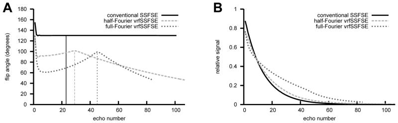
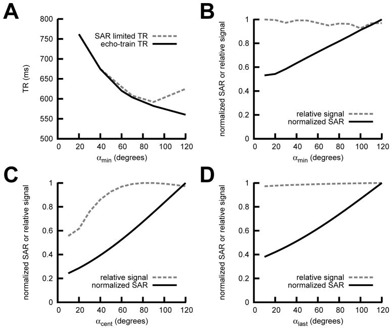
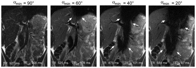
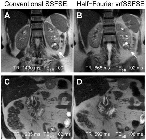
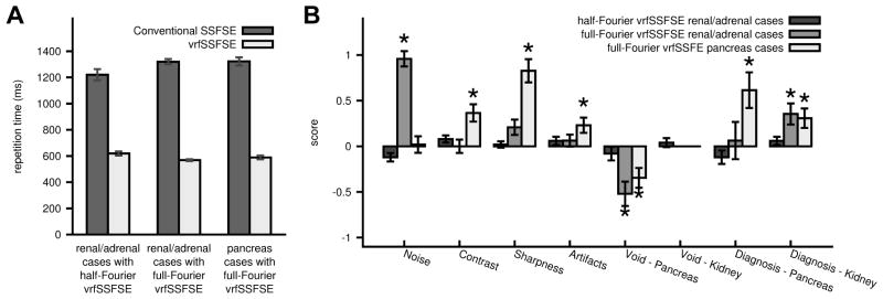
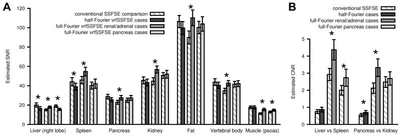
Materials and methods: A variable refocusing flip angle SSFSE sequence (vrfSSFSE) was designed and implemented, with simulations and volunteer scans performed to determine suitable flip angle modulation parameters. With Institutional Review Board (IRB) approval/informed consent, patients referred for 3T abdominal magnetic resonance imaging (MRI) were scanned with conventional SSFSE and either half-Fourier (n = 25) or full-Fourier vrfSSFSE (n = 50). Two blinded radiologists semiquantitatively scored images on a scale from -2 to 2 for contrast, noise, sharpness, artifacts, cardiac motion-related signal loss, and the ability to evaluate the pancreas and kidneys.

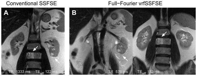
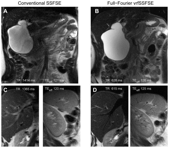
Results: vrfSSFSE demonstrated significantly increased speed (∼2-fold, P < 0.0001). Significant improvements in image quality parameters with full-Fourier vrfSSFSE included increased contrast, sharpness, and visualization of pancreatic and renal structures with higher bandwidth technique (mean scores 0.37, 0.83, 0.62, and 0.31, respectively, P ≤ 0.001), and decreased image noise and improved visualization of renal structures when used with an equal bandwidth technique (mean scores 0.96 and 0.35, respectively, P < 0.001). Increased cardiac motion-related signal loss with full-Fourier vrfSSFSE was seen in the pancreas but not the kidney.

Conclusion: vrfSSFSE increases speed at 3T over conventional SSFSE via reduced SAR, and when combined with full-Fourier acquisition can improve image quality, although with some increased sensitivity to cardiac motion-related signal loss.

Keywords: echo stabilization; single shot fast spin echo; specific absorption rate; variable refocusing flip angle.


Figures

Figure 1
Figure 1
(A) Example flip angle trains for conventional SSFSE (α = 130°), half-Fourier vrfSSFSE (αinit = 130°, αmin = 90°, αcent = 100°, αlast = 45°), and full-Fourier vrfSSFSE (αinit = 130°, αmin = 60°, αcent = 100°, αlast = 45°). The vertical lines indicate where the center of k-space is traversed. Echo trains were generated to achieve an effective TE of 130 ms. The full-Fourier vrfSSFSE example has a shorter echo train as a smaller number of phase encodes were utilized in this example. (B) Corresponding simulated signal curves for the different echo trains, note the flattening of the signal curve for echo trains with decreased values of αmin.
Figure 2
Figure 2
Half-Fourier vrfSSFSE EPG Simulation Results. (A) Echo train length limited TR and specific absorption rate (SAR) limited TR vs. αmin for TE 100 ms. Normalized signal output and normalized SAR for (B) αmin, (C) αcent, and (D) αlast. The non-varying parameters were αinit=130°, αmin=90°, αcent=100°, and αlast=45°.
Figure 3
Figure 3
Cardiac motion related signal loss for full-Fourier vrfSSFSE sequences with varying αmin. Representative slices are shown for the most severely affected slices, although this artifact (arrows) is intermittent presumably related to timing of acquisition relative to phase of the cardiac cycle. For higher αmin (e.g. 90°) and full-Fourier technique only a relatively long effective TE is obtainable (> 180 ms) resulting in low signal images. For lower αmin (e.g. < 50°) the artifact becomes overly problematic and compromises the diagnostic ability of the images.
Figure 4
Figure 4
Examples of conventional SSFSE compared to half-Fourier vrfSSFSE. (A) Conventional SSFSE and (B) half-Fourier vrfSSFSE images from a patient with renal cell carcinoma of the left kidney (arrow). (C) Conventional SSFSE and (D) half-Fourier vrfSSFSE images from a patient with a mildly dilated pancreatic and common bile duct. Images are mildly cropped from the full field of view. Note the equivalent image quality.
Figure 5
Figure 5
Comparison of conventional SSFSE and vrfSSFSE. A: Comparison of repetition times (TR). For each pair (conventional SSFSE versus vrfSSFSE) the differences were significant (p<0.0001). B: Results of semi-quantitative grading for noise, contrast, sharpness, general artifacts, cardiac motion related signal loss over the pancreas and kidney, and the ability to diagnose pancreatic or renal abnormalities. * indicates significant differences. The scoring system utilized is explained in Table 2. Negative numbers favor conventional SSFSE, positive numbers favor vrfSSFSE. The presence/absence of a cardiac motion related signal void impacting the kidneys was not statistically evaluated in the full-Fourier vrfSSFSE cases, as it was not seen in any of the cases. Error bars are standard error of the mean.
Figure 6
Figure 6
Estimated signal to noise (SNR) and contrast to noise (CNR) based on clinical data. (A) SNR and (B) CNR are shown for half-Fourier vrfSSFSE renal/adrenal, full-Fourier vrfSSFSE renal/adrenal, and full-Fourier pancreas cases, adjacent to their comparison conventional SSFSE. SNR was calculated as mean signal in the tissue ROI divided by standard deviation from an ROI placed over extra-corporeal air. CNR was calculated as the absolute difference in signal for the two tissues divided by the standard deviation from an ROI placed over extra-corporeal air. * indicates significant differences between the conventional SSFSE and the corresponding vrfSSFSE sequence. Error bars are standard error of the mean.
Figure 7
Figure 7
Comparison of conventional SSFSE to full-Fourier vrfSSFSE for imaging of the kidney. (A) Conventional SSFSE image showing a renal cell carcinoma of the left kidney (right arrow) and an L3 vertebral body lesion that proved to be a hemangioma (left arrow). (B) Two images from the same full-Fourier vrfSSFSE acquisition showing the small renal cell carcinoma (left image) and the hemangioma (right image). Note the decreased noise compared to the conventional SSFSE image, and improved visualization of the characteristic vertical striations of the L3 vertebral body hemangioma. The differences in positioning are due to the conventional SSFSE needing to be acquired using respiratory triggering (images acquired at end-expiration). As the vrfSSFSE images can be acquired in half the time, the full stack of 38 images was acquired during a single 22 s breath hold. Images are moderately cropped.
Figure 8
Figure 8
Comparison of conventional SSFSE to full-Fourier vrfSSFSE acquired for evaluation of the pancreas and biliary ducts. (A) Conventional SSFSE and (B) full-Fourier vrfSSFSE images demonstrate a type I choledochocele that causes mass effect upon the pancreas as seen by the altered course of the pancreatic duct. Images are mildly cropped. Close up views from a different patient of the liver and spleen for (C) conventional SSFSE and (D) full-Fourier vrfSSFSE highlight the more prominent black blood appearance of vasculature with the vrfSSFSE technique. This is best seen in the hepatic vein in the liver and small branching vessels radiating from the hilum of the spleen.

References

    1. Chavhan GB, Babyn PS, Vasanawala SS. Abdominal MR imaging in children: motion compensation, sequence optimization, and protocol organization. Radiographics. 2013;33:703–719. - PubMed
    1. Yang RK, Roth CG, Ward RJ, deJesus JO, Mitchell DG. Optimizing abdominal MR imaging: approaches to common problems. Radiographics. 2010:185–199. - PubMed
    1. Bhosale P, Ma J, Choi H. Utility of the FIESTA pulse sequence in body oncologic imaging: review. AJR Am J Roentgenol. 2009;192:S83–93. - PubMed
    1. Hennig J, Weigel M, Scheffler K. Multiecho sequences with variable refocusing flip angles: optimization of signal behavior using smooth transitions between pseudo steady states (TRAPS) Magn Reson Med. 2003;49:527–535. - PubMed
    1. Madhuranthakam AJ, Busse RF, Brittain JH, Rofsky NM, Alsop DC. Sensitivity of low flip angle SSFSE of the abdomen to cardiac motion. Proceedings of the 15th Annual Meeting of ISMRM; Berlin, Germany. 2007; p. abstract 2523.