. 2015 Jul 7;23(7):1179-89.
doi: 10.1016/j.str.2015.04.022. Epub 2015 Jun 18.

Light Signaling Mechanism of Two Tandem Bacteriophytochromes

Affiliations

Light Signaling Mechanism of Two Tandem Bacteriophytochromes

Xiaojing Yang et al. Structure. .

Abstract

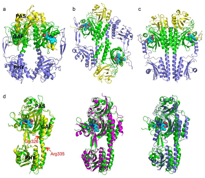
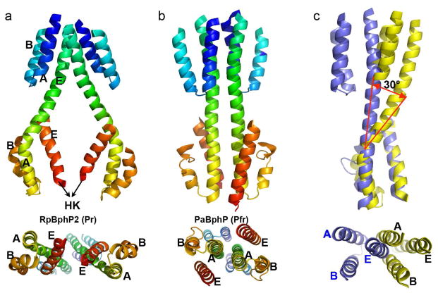
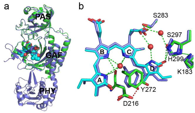
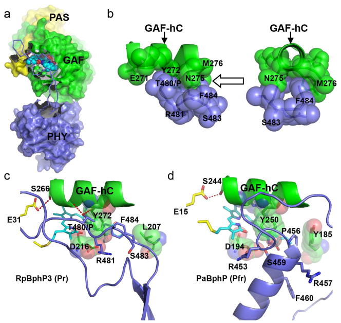
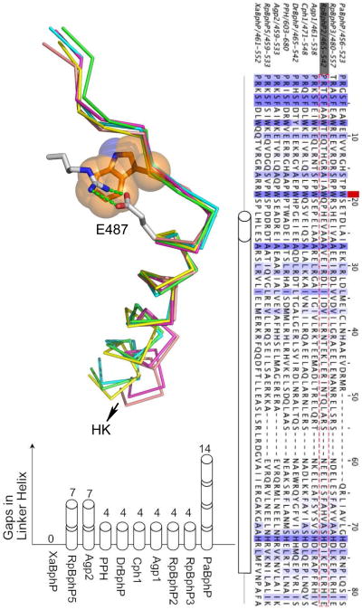
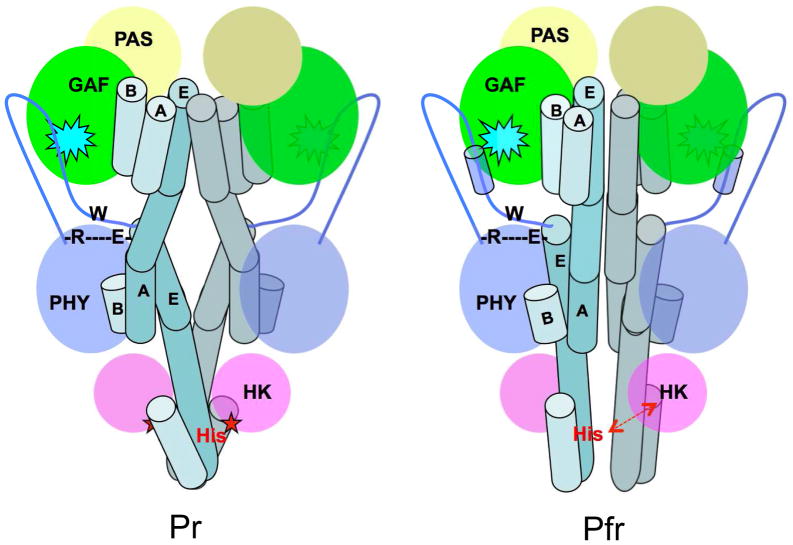
RpBphP2 and RpBphP3, two tandem bacteriophytochromes from the photosynthetic bacterium Rhodopseudomonas palustris, share high sequence identity but exhibit distinct photoconversion behavior. Unlike the canonical RpBphP2, RpBphP3 photoconverts to an unusual near-red-absorbing (Pnr) state; both are required for synthesis of light-harvesting complexes under low-light conditions. Here we report the crystal structures of the photosensory core modules of RpBphP2 and RpBphP3. Despite different quaternary structures, RpBphP2 and RpBphP3 adopt nearly identical tertiary structures. The RpBphP3 structure reveals tongue-and-groove interactions at the interface between the GAF and PHY domains. A single mutation in the PRxSF motif at the GAF-PHY interface abolishes light-induced formation of the Pnr state in RpBphP3, possibly due to altered structural rigidity of the chromophore-binding pocket. Structural comparisons suggest that long-range signaling involves structural rearrangement of the helical spine at the dimer interface. These structures, together with mutational studies, provide insights into photoconversion and the long-range signaling mechanism in phytochromes.


Figures

Fig. 1
Fig. 1. Crystal structures of RpBphP2 and RpBphP3
a) Ribbon diagram of the parallel dimer of RpBphP2-Ctag in the Pr state. The PAS (yellow), GAF (green) and PHY (blue) domains are juxtaposed along the dimer interface, where a large void is found. The BV chromophores are shown in cyan spheres. b) Ribbon diagram of the anti-parallel dimer of RpBphP3 in the Pr state. c) Parallel dimer structure of PaBphP in the Pfr state exhibits a tighter dimer interface (PDB ID: 3NHQ). d) Pairwise comparison of the tertiary structures of BphP monomers: RpBphP3 (green; as reference); RpBphP2 (yellow), Cph1 (magenta) and PaBphP (blue). The red arrows mark the locations of kink at the GAF-PHY linker helix (RpBphP3 numbering). Related to Figure S1 and Figure S2a.
Fig. 2
Fig. 2. Helices at the dimer interface
Interfacial helices in the parallel dimer structures of RpBphP2 (a) and PaBphP (b) are shown in rainbow colors from blue to red corresponding to the residue order from the N- to C- termini. Equivalent helices A, B and E in the GAF and PHY domains are labeled. Lower panels represent the bottom views of the PCM dimer, as seen from the HK domain. c) Structural alignment of interfacial helices in RpBphP2 (yellow) and PaBphP (blue) according to the 3-helix bundle of the PHY domain. The lower panel represents the top view of the superposition.
Figure 3
Figure 3. Structural comparisons between the CBD and PCM constructs of RpBphP3
a) Superposition of the RpBphP3-PCM (blue) and RpBphP3-CBD (green/cyan) monomers shows nearly identical tertiary structures in the PAS and GAF domains; b) Structural differences in the chromophore-binding pocket between PCM (cyan and green) and CBD (blue) of RpBphP3. Dashed green lines represent hydrogen bonds. Water molecules are marked in red spheres. Related to Figure S1c.
Figure 4
Figure 4. The GAF-PHY interface
a) In the Pr structure of RpBphP3-PCM, a long extended arm spanning residues 454-494 (in ribbon representation) protruding from the core of the PHY domain (blue) shields a large surface area of the GAF domain (green) near the chromophore (in cyan). b) “Tongue-and-groove” contacts between the side chains of the GAF-hC helix (green) and the PRxSF motif (blue) in two orthogonal views as indicated by the arrow. c) In RpBphP3-PCM in the Pr state, the PRxSF motif adopts an extended conformation, in which Thr480 in place of Pro makes direct interactions with the GAF-hC helix (hydrogen bonds are marked in red dashed lines). Residues Tyr272, Asp216 and Leu207 in the GAF domain are shown in spheres. d) In PaBphP in the Pfr state, the PRxSF motif is part of a 3-turn helix, in which side chains adopt very different orientations relative to the GAF-hC helix than in RpBphP3-PCM. Related to Figure S3 and Figure S5.
Fig. 5
Fig. 5. The PHY-HK linker helix
a) Superposition of five crystal structures in the junction region between the arm of the PHY domain and the HK domain. RpBphP2 (PDB 4R6L; green), RpBphP3 (PDB 4R70; yellow), Cph1 (2VEA; magenta), PaBphP (3C2W; blue) and RpBphP1 (4EH0; orange). The side chain of the conserved Trp residue (Trp483 of RpBphP2) is shown in orange, and the side chains of Arg439 and Glu487 are in grey with stick. Green dashed lines mark the salt bridges. b) Sequence alignment between the PRxSF motif and the phospho-accepting histidine in 10 representative BphPs. The red square marks the position of the conserved Trp483 in RpBphP2 (in gray box and sequence is highlighted red dashed). Gaps are filled by short dashes. A long tube represents the continuous PHY-HK linker helix according to the secondary structure prediction. The length of the short tube measures one α-helical turn. c) Length differences in the PHY-HK linker helices are represented by gaps in the sequence alignment as measured by the number of helical turns. Related to Figure S4.
Fig. 6
Fig. 6. Long-range signaling mechanism in BphPs
Schematic illustration of the BphP parallel dimer structures in the Pr (left) and Pfr (right) states. Important structural elements involved in long-range signaling are highlighted: the BV chromophore embedded in the GAF domain (cyan spike), the arm (blue line with the tube representing a helix) of the PHY domain, the R-W-E/D motif, and the phospho-accepting histidine residue (red), and the long linker helices (in teal tubes). Interfacial helices in one monomer are labeled according to the crystal structures of RpBphP2-PCM (Pr) and PaBphP-PCM (Pfr). The core of each modular domain is colored as the following: PAS (yellow), GAF (green), PHY (blue) and HK (magenta). Straightening of the linker helices in the helical spine affects relative positioning of the catalytic ATPase (magenta oval) and His. Specifically, activation of HK auto-phosphorylation is marked by a red star when ATPase is sufficiently close to His. Deactivation of HK is indicated with a red double-arrow as His separates from the active site of ATPase.

References

    1. Adams PD, Afonine PV, Bunkóczi G, Chen VB, Davis IW, Echols N, Headd JJ, Hung LW, Kapral GJ, Grosse-Kunstleve RW, et al. PHENIX: a comprehensive Python-based system for macromolecular structure solution. Acta Crystallogr D Biol Crystallogr. 2010;66:213–221. - PMC - PubMed
    1. Anantharaman V, Balaji S, Aravind L. The signaling helix: a common functional theme in diverse signaling proteins. Biol Direct. 2006;1:25. - PMC - PubMed
    1. Anders K, Daminelli-Widany G, Mroginski MA, von Stetten D, Essen LO. Structure of the Cyanobacterial Phytochrome 2 Photosensor Implies a Tryptophan Switch for Phytochrome Signaling. J Biol Chem. 2013;288:35714–35725. - PMC - PubMed
    1. Ayers RA, Moffat K. Changes in Quaternary Structure in the Signaling Mechanisms of PAS Domains. Biochemistry (Mosc) 2008;47:12078–12086. - PMC - PubMed
    1. Bellini D, Papiz MZ. Structure of a Bacteriophytochrome and Light-Stimulated Protomer Swapping with a Gene Repressor. Structure. 2012a;20:1436–1446. - PubMed

Publication types

LinkOut - more resources