Review
. 2016 Jan;22(1):33-8.
doi: 10.1111/odi.12353. Epub 2015 Jul 2.

Mouse genetic models for temporomandibular joint development and disorders

Affiliations
Review

Mouse genetic models for temporomandibular joint development and disorders

A Suzuki et al. Oral Dis. 2016 Jan.

Abstract

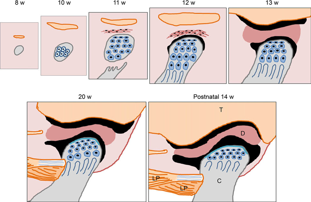
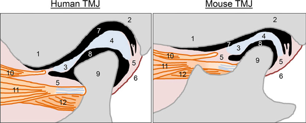
The temporomandibular joint (TMJ) is a synovial joint essential for hinge and sliding movements of the mammalian jaw. Temporomandibular joint disorders (TMD) are dysregulations of the muscles or the TMJ in structure, function, and physiology, and result in pain, limited mandibular mobility, and TMJ noise and clicking. Although approximately 40-70% adults in the USA have at least one sign of TMD, the etiology of TMD remains largely unknown. Here, we highlight recent advances in our understanding of TMD in mouse models.

Keywords: bones and joints; craniofacial; developmental disorders; genetics; mouse model; temporomandibular joint disorders.


Conflict of interest statement

Conflict of interest

None declared.

Figures

Figure 1
Figure 1
Time course of temporomandibular joint development in humans. At the 8th week of gestation, mesenchymal condensation forms at the TMJ region. Orange, mesenchymal condensation of the future temporalis bone; gray, mesenchymal condensation of the future mandibular condyle. At the 10th week of gestation, mesenchymal cells differentiate into chondrocytes and start to form the core of cartilage which is the future mandibular condyle. The temporal bone starts intramembranous ossification. During the 11th week of gestation, intramembranous ossification of the ramus of the mandible reaches the base of the future condyle. There are no joint cavities during this stage. The mesenchymal condensation that forms the anlage of the articular disk appears in connective tissue between the anlage of the temporal bone and the mandibular condyle. During the 12th week of gestation, a small space or cleft appears between the anlage of the articular disk and the mandibular condyle that defines the initial formation of the lower articular cavity. During the 13th week of gestation, the organization of the upper articular cavity starts between the temporal bone and the articular disk. At approximately 20th week of gestation, the glenoid fossa of the temporal bone forms, but the articular surface of the temporal bone has a flat surface. At postnatal 14th week, the glenoid fossa and the articular eminence of the temporal bone well form and fit with the shape of the mandibular condyle. T: the temporal bone, D: the articular disk, C: the mandibular condyle, LP: the lateral pterygoid muscle
Figure 2
Figure 2
Comparison of the structure of the temporomandibular joint between humans and mice. TMJ, temporomandibular joint. 1: the articular eminence of the temporal bone, 2: the glenoid fossa of the temporal bone, 3: anterior band of the articular disk, 4: posterior band of the articular disk, 5: connective tissue, 6: the posterior joint capsule, 7: the upper articular cavity, 8: the lower articular cavity, 9: mandibular condyle, 10: a part of upper head of the lateral pterygoid muscle, associated with the articular disk, 11: upper head of the lateral pterygoid muscle, connected with the mandibular condyle, 12: lower head of the lateral pterygoid muscle, connected with the mandibular condyle

Similar articles

Cited by

References

    1. Benjamin M, Ralphs JR. Biology of fibrocartilage cells. Int Rev Cytol. 2004;233:1–45. - PubMed
    1. Gu S, Wei N, Yu L, Fei J, Chen Y. Shox2-deficiency leads to dysplasia and ankylosis of the temporomandibular joint in mice. Mech Dev. 2008;125:729–742. - PMC - PubMed
    1. Gu S, Wu W, Liu C, et al. BMPRIA mediated signaling is essential for temporomandibular joint development in mice. PLoS ONE. 2014;9:e101000. - PMC - PubMed
    1. Herring SW. TMJ anatomy and animal models. J Musculoskelet Neuronal Interact. 2003;3:391–394. discussion 406-7. - PMC - PubMed
    1. Hinton RJ. Genes that regulate morphogenesis and growth of the temporomandibular joint: a review. Dev Dyn. 2014;243:864–874. - PubMed

Publication types

Substances